")
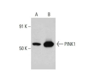
: sc-517353. Western blot analysis of PINK1 expression in A-431 (A) and Sol8 (B) whole cell lysates.")
PINK1 Antibody (38CT20.8.5): sc-517353
- PINK1 Antibody (38CT20.8.5) is a mouse monoclonal IgG1 κ PINK1 antibody, cited in 79 publications, provided at 50 µg/0.5 ml
- raised against a recombinant protein corresponding to PINK1 of human origin
- PINK1 Antibody (38CT20.8.5) is recommended for detection of PINK1 of mouse, rat and human origin by WB, IP, IF and IHC(P)
- m-IgG Fc BP-HRP and m-IgG1 BP-HRP are the preferred secondary detection reagents for PINK1 Antibody (38CT20.8.5) for WB and IHC(P) applications. These reagents are now offered in bundles with PINK1 Antibody (38CT20.8.5) (see ordering information below).

QUICK LINKS
SEE ALSO...
PINK1 Antibody (38CT20.8.5) is a mouse monoclonal IgG1 kappa light chain antibody that detects PINK1 protein of mouse, rat, and human origin by western blotting (WB), immunoprecipitation (IP), immunofluorescence (IF), and immunohistochemistry with paraffin-embedded sections (IHCP). anti-PINK1 antibody (38CT20.8.5) is available as a non-conjugated monoclonal isotype antibody. PINK1, or PTEN induced putative kinase 1, is a crucial member of the serine/threonine protein kinase family and functions as a tumor suppressor. PINK1 is primarily localized in mitochondria, where PINK1 plays a vital role in maintaining mitochondrial integrity and function. PINK1′s presence in mitochondria helps protect cells from mitochondrial dysfunction, particularly during cellular stress, by phosphorylating mitochondrial proteins essential for energy production and apoptosis regulation. Mutations in the PINK1 gene (PARK6) can lead to altered autophosphorylation activity, which is linked to early onset Parkinson′s disease, a neurodegenerative disorder characterized by motor symptoms such as resting tremor and rigidity. PINK1′s widespread expression in tissues such as testis, skeletal muscle, and heart underscores PINK1′s importance in various physiological processes, while lower expression in tissues like pancreas and brain highlights specialized roles in cellular health and disease.
Alexa Fluor® is a trademark of Molecular Probes Inc., OR., USA
LI-COR® and Odyssey® are registered trademarks of LI-COR Biosciences
PINK1 Antibody (38CT20.8.5) References:
- Growth-suppressive effects of BPOZ and EGR2, two genes involved in the PTEN signaling pathway. | Unoki, M. and Nakamura, Y. 2001. Oncogene. 20: 4457-65. PMID: 11494141
- Hereditary early-onset Parkinson's disease caused by mutations in PINK1. | Valente, EM., et al. 2004. Science. 304: 1158-60. PMID: 15087508
- The gene responsible for PARK6 Parkinson's disease, PINK1, does not influence common forms of parkinsonism. | Healy, DG., et al. 2004. Ann Neurol. 56: 329-35. PMID: 15349859
- Novel PINK1 mutations in early-onset parkinsonism. | Hatano, Y., et al. 2004. Ann Neurol. 56: 424-7. PMID: 15349870
- Analysis of the PINK1 gene in a large cohort of cases with Parkinson disease. | Rogaeva, E., et al. 2004. Arch Neurol. 61: 1898-904. PMID: 15596610
- Mitochondrial import and enzymatic activity of PINK1 mutants associated to recessive parkinsonism. | Silvestri, L., et al. 2005. Hum Mol Genet. 14: 3477-92. PMID: 16207731
- Analysis of the PINK1 gene in a cohort of patients with sporadic early-onset parkinsonism in Taiwan. | Fung, HC., et al. 2006. Neurosci Lett. 394: 33-6. PMID: 16257123
- Loss of PINK1 function promotes mitophagy through effects on oxidative stress and mitochondrial fission. | Dagda, RK., et al. 2009. J Biol Chem. 284: 13843-13855. PMID: 19279012
- PINK1 phosphorylates Drp1S616 to regulate mitophagy-independent mitochondrial dynamics. | Han, H., et al. 2020. EMBO Rep. 21: e48686. PMID: 32484300
- Targeting ATAD3A-PINK1-mitophagy axis overcomes chemoimmunotherapy resistance by redirecting PD-L1 to mitochondria. | Xie, XQ., et al. 2023. Cell Res. 33: 215-228. PMID: 36627348
- Unconventional initiation of PINK1/Parkin mitophagy by Optineurin. | Nguyen, TN., et al. 2023. Mol Cell. 83: 1693-1709.e9. PMID: 37207627
- PINK1 and Parkin regulate IP3R-mediated ER calcium release. | Ham, SJ., et al. 2023. Nat Commun. 14: 5202. PMID: 37626046
Ordering Information
| Product Name | Catalog # | UNIT | Price | Qty | FAVORITES | |
PINK1 Antibody (38CT20.8.5) | sc-517353 | 50 µg/0.5 ml | $322.00 | |||
PINK1 Antibody (38CT20.8.5): m-IgG Fc BP-HRP Bundle | sc-541103 | 50 µg Ab; 10 µg BP | $361.00 | |||
PINK1 Antibody (38CT20.8.5): m-IgG1 BP-HRP Bundle | sc-542582 | 50 µg Ab; 20 µg BP | $361.00 |