")
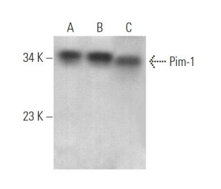
: sc-13513. Western blot analysis of Pim-1 expression in HUV-EC-C (A), Raji (B) and M1 (C) whole cell lysates.")
: sc-13513. Western blot analysis of Pim-1 expression in MEG-01 whole cell lysate.")
: sc-13513. Western blot analysis of Pim-1 expression in K-562 whole cell lysate.")
Pim-1 Antibody (12H8): sc-13513
- Pim-1 Antibody (12H8) is a mouse monoclonal IgG1 κ, cited in 99 publications, provided at 200 µg/ml
- raised against full length of Pim-1 of mouse origin
- Anti-Pim-1 Antibody (12H8) is recommended for detection of Pim-1 of mouse, rat and human origin by WB and IP
- Pim-1 Antibody (12H8) is available conjugated to Alexa Fluor® 546 or Alexa Fluor® 594 for WB (RGB), IF, IHC(P) and FCM
- also available conjugated to Alexa Fluor® 680 or Alexa Fluor® 790 for WB (NIR), IF and FCM; for use with Near-Infrared (NIR) detection systems, such as LI-COR®Odyssey®, iBright™ FL1000, FluorChem™, Typhoon, Azure and other comparable systems
- m-IgG Fc BP-HRP and m-IgG1 BP-HRP are the preferred secondary detection reagents for Pim-1 Antibody (12H8) for WB applications. These reagents are now offered in bundles with Pim-1 Antibody (12H8) (see ordering information below).

QUICK LINKS
Pim-1 Antibody (12H8) is a mouse monoclonal IgG1 kappa light chain antibody that detects Pim-1 in mouse, rat, and human samples through western blotting (WB) and immunoprecipitation (IP). Pim-1 (12H8) antibody is available in both conjugated and non-conjugated forms, providing versatility for various experimental needs. Pim-1 is a serine/threonine kinase that plays a crucial role in cell signaling pathways, particularly in lymphoid cell transformation, where Pim-1 collaborates with c-Myc to promote cell proliferation and survival. Pim-1 is predominantly located in the cytoplasm and nucleus, which is significant as Pim-1 localization is essential for function in regulating cell cycle and apoptosis. Specifically, Pim-1 expression increases during transition from early to late G1 phase, remaining elevated at G1/S boundary and G2 phases, thereby influencing cell cycle progression. Additionally, Pim-1 is regulated at both transcriptional and translational levels, with Pim-1 expression being induced by interleukin-2 stimulation, highlighting Pim-1′s importance in T cell differentiation and immune responses. Furthermore, Pim-1 has been shown to stimulate c-Myc-mediated apoptosis upstream of caspase-3-like proteases, underscoring Pim-1′s role in maintaining cellular homeostasis and preventing uncontrolled cell growth.
Alexa Fluor® is a trademark of Molecular Probes Inc., OR., USA
LI-COR® and Odyssey® are registered trademarks of LI-COR Biosciences
Pim-1 Antibody (12H8) References:
- Understanding the functional discrepancy of Pim-1 in cancer. | Ouhtit, A., et al. 2015. Front Biosci (Elite Ed). 7: 208-14. PMID: 25553374
- PIM-1 may function as an oncogene in cervical cancer via activating the EGFR signaling. | Yang, H., et al. 2020. Int J Biol Markers. 35: 67-73. PMID: 32914663
- Pim-1 kinase is a positive feedback regulator of the senescent lung fibroblast inflammatory secretome. | Gao, AY., et al. 2022. Am J Physiol Lung Cell Mol Physiol. 323: L685-L697. PMID: 36223640
- Targeting macrophagic PIM-1 alleviates osteoarthritis by inhibiting NLRP3 inflammasome activation via suppressing mitochondrial ROS/Cl- efflux signaling pathway. | Zhang, Z., et al. 2023. J Transl Med. 21: 452. PMID: 37422640
- Inhibition of DYRK1A attenuates vascular remodeling in pulmonary arterial hypertension via suppressing STAT3/Pim-1/NFAT pathway. | Lan, C., et al. 2024. Clin Exp Hypertens. 46: 2297642. PMID: 38147409
- The role of Pim-1 kinases in inflammatory signaling pathways. | Baek, HS., et al. 2024. Inflamm Res. 73: 1671-1685. PMID: 39079978
- Ubiquitous expression and cell cycle regulation of the protein kinase PIM-1. | Liang, H., et al. 1996. Arch Biochem Biophys. 330: 259-65. PMID: 8660654
- The effect of IL-2 treatment on transcriptional attenuation in proto-oncogenes pim-1 and c-myb in human thymic blast cells. | Rohwer, F., et al. 1996. J Immunol. 157: 643-9. PMID: 8752912
- Pim-1 kinase stimulates c-Myc-mediated death signaling upstream of caspase-3 (CPP32)-like protease activation. | Mochizuki, T., et al. 1997. Oncogene. 15: 1471-80. PMID: 9333023
- Pim-1 protein expression is regulated by its 5'-untranslated region and translation initiation factor elF-4E. | Hoover, DS., et al. 1997. Cell Growth Differ. 8: 1371-80. PMID: 9419425
- Evidence implicating Gfi-1 and Pim-1 in pre-T-cell differentiation steps associated with beta-selection. | Schmidt, T., et al. 1998. EMBO J. 17: 5349-59. PMID: 9736613
- Pim-1 kinase and p100 cooperate to enhance c-Myb activity. | Leverson, JD., et al. 1998. Mol Cell. 2: 417-25. PMID: 9809063
Ordering Information
| Product Name | Catalog # | UNIT | Price | Qty | FAVORITES | |
Pim-1 Antibody (12H8) | sc-13513 | 200 µg/ml | $322.00 | |||
Pim-1 Antibody (12H8): m-IgG Fc BP-HRP Bundle | sc-526549 | 200 µg Ab; 10 µg BP | $361.00 | |||
Pim-1 Antibody (12H8): m-IgG1 BP-HRP Bundle | sc-531922 | 200 µg Ab; 20 µg BP | $361.00 | |||
Pim-1 Antibody (12H8) Alexa Fluor® 546 | sc-13513 AF546 | 200 µg/ml | $364.00 | |||
Pim-1 Antibody (12H8) Alexa Fluor® 594 | sc-13513 AF594 | 200 µg/ml | $364.00 | |||
Pim-1 Antibody (12H8) Alexa Fluor® 680 | sc-13513 AF680 | 200 µg/ml | $364.00 | |||
Pim-1 Antibody (12H8) Alexa Fluor® 790 | sc-13513 AF790 | 200 µg/ml | $364.00 |