")
PEA3 Antibody (G-10): sc-166629
- PEA3 Antibody (G-10) is a mouse monoclonal IgG1 κ PEA3 antibody, cited in 9 publications, provided at 200 µg/ml
- raised against amino acids 171-290 of PEA3 of human origin
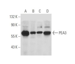
- PEA3 Antibody (G-10) is recommended for detection of PEA3 of mouse, rat and human origin by WB, IP, IF and ELISA
- Anti-PEA3 Antibody (G-10) is available conjugated to agarose for IP; HRP for WB, IHC(P) and ELISA; and to either phycoerythrin or FITC for IF, IHC(P) and FCM
- also available conjugated to Alexa Fluor® 488, Alexa Fluor® 546, Alexa Fluor® 594 or Alexa Fluor® 647 for WB (RGB), IF, IHC(P) and FCM, and for use with RGB fluorescent imaging systems, such as iBright™ FL1000, FluorChem™, Typhoon, Azure and other comparable systems
- also available conjugated to Alexa Fluor® 680 or Alexa Fluor® 790 for WB (NIR), IF and FCM; for use with Near-Infrared (NIR) detection systems, such as LI-COR®Odyssey®, iBright™ FL1000, FluorChem™, Typhoon, Azure and other comparable systems
- TransCruz reagent for Gel Supershift and ChIP applications (sc-166629 X, 200 µg/0.1 ml)
- m-IgG Fc BP-HRP, m-IgG1 BP-HRP and m-IgGκ BP-HRP are the preferred secondary detection reagents for PEA3 Antibody (G-10) for WB applications. These reagents are now offered in bundles with PEA3 Antibody (G-10) (see ordering information below).

QUICK LINKS
PEA3 Antibody (G-10) is a mouse monoclonal IgG1 kappa light chain antibody that detects PEA3 protein of mouse, rat, and human origin by western blotting (WB), immunoprecipitation (IP), immunofluorescence (IF), and enzyme-linked immunosorbent assay (ELISA). PEA3 Antibody (G-10) is available in both non-conjugated and various conjugated forms, including agarose, horseradish peroxidase (HRP), phycoerythrin (PE), fluorescein isothiocyanate (FITC), and multiple Alexa Fluor® conjugates. PEA3, also known as ETS Variant Transcription Factor 4, plays a crucial role in regulating gene expression during development and cellular differentiation, particularly in epithelial and fibroblastic cells. PEA3′s ability to bind specific DNA motifs, such as the PEA-3 motif (5′-AGGAAG-3′), allows PEA3 to influence the transcription of target genes, which is vital for processes like cell proliferation and migration. Notably, PEA3 is not expressed in hematopoietic cells, distinguishing PEA3 from other Ets family members like Ets-1 and Ets-2, which are primarily found in blood cell lineages. This specificity in expression highlights the unique functional roles of Ets proteins in different cell types and developmental contexts, making PEA3 a significant marker for studying epithelial and fibroblast biology.
Alexa Fluor® is a trademark of Molecular Probes Inc., OR., USA
LI-COR® and Odyssey® are registered trademarks of LI-COR Biosciences
PEA3 Antibody (G-10) References:
- The pea3 subfamily ets genes are required for HER2/Neu-mediated mammary oncogenesis. | Shepherd, TG., et al. 2001. Curr Biol. 11: 1739-48. PMID: 11719215
- The Pea3 Ets transcription factor regulates differentiation of multipotent progenitor cells during mammary gland development. | Kurpios, NA., et al. 2009. Dev Biol. 325: 106-21. PMID: 18977342
- Pea3 transcription factors and wnt1-induced mouse mammary neoplasia. | Baker, R., et al. 2010. PLoS One. 5: e8854. PMID: 20107508
- PEA3/ETV4-related transcription factors coupled with active ERK signalling are associated with poor prognosis in gastric adenocarcinoma. | Keld, R., et al. 2011. Br J Cancer. 105: 124-30. PMID: 21673681
- NOTCH-1 and NOTCH-4 are novel gene targets of PEA3 in breast cancer: novel therapeutic implications. | Clementz, AG., et al. 2011. Breast Cancer Res. 13: R63. PMID: 21679465
- The role of Pea3 group transcription factors in esophageal squamous cell carcinoma. | Yuen, HF., et al. 2011. Am J Pathol. 179: 992-1003. PMID: 21689625
- PEA3 activates CXCR4 transcription in MDA-MB-231 and MCF7 breast cancer cells. | Gu, S., et al. 2011. Acta Biochim Biophys Sin (Shanghai). 43: 771-8. PMID: 21831961
- A positive role of DBC1 in PEA3-mediated progression of estrogen receptor-negative breast cancer. | Kim, HJ., et al. 2015. Oncogene. 34: 4500-8. PMID: 25417701
- Pea3 determines the isthmus region at the downstream of Fgf8-Ras-ERK signaling pathway. | Harada, H., et al. 2015. Dev Growth Differ. 57: 657-66. PMID: 26691276
- Prognostic value of PEA3 subfamily gene expression in cholangiocarcinoma. | Wang, L., et al. 2024. World J Gastrointest Oncol. 16: 4014-4027. PMID: 39350976
Ordering Information
| Product Name | Catalog # | UNIT | Price | Qty | FAVORITES | |
PEA3 Antibody (G-10) | sc-166629 | 200 µg/ml | $322.00 | |||
PEA3 Antibody (G-10): m-IgG Fc BP-HRP Bundle | sc-529010 | 200 µg Ab; 10 µg BP | $361.00 | |||
PEA3 Antibody (G-10): m-IgGκ BP-HRP Bundle | sc-521681 | 200 µg Ab, 40 µg BP | $361.00 | |||
PEA3 Antibody (G-10): m-IgG1 BP-HRP Bundle | sc-543266 | 200 µg Ab; 20 µg BP | $361.00 | |||
PEA3 Antibody (G-10) X | sc-166629 X | 200 µg/0.1 ml | $322.00 | |||
PEA3 Antibody (G-10) AC | sc-166629 AC | 500 µg/ml, 25% agarose | $424.00 | |||
PEA3 Antibody (G-10) HRP | sc-166629 HRP | 200 µg/ml | $322.00 | |||
PEA3 Antibody (G-10) FITC | sc-166629 FITC | 200 µg/ml | $336.00 | |||
PEA3 Antibody (G-10) PE | sc-166629 PE | 200 µg/ml | $349.00 | |||
PEA3 Antibody (G-10) Alexa Fluor® 488 | sc-166629 AF488 | 200 µg/ml | $364.00 | |||
PEA3 Antibody (G-10) Alexa Fluor® 546 | sc-166629 AF546 | 200 µg/ml | $364.00 | |||
PEA3 Antibody (G-10) Alexa Fluor® 594 | sc-166629 AF594 | 200 µg/ml | $364.00 | |||
PEA3 Antibody (G-10) Alexa Fluor® 647 | sc-166629 AF647 | 200 µg/ml | $364.00 | |||
PEA3 Antibody (G-10) Alexa Fluor® 680 | sc-166629 AF680 | 200 µg/ml | $364.00 | |||
PEA3 Antibody (G-10) Alexa Fluor® 790 | sc-166629 AF790 | 200 µg/ml | $364.00 |