")
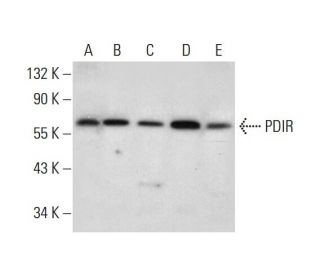
: sc-390862. Western blot analysis of PDIR expression in HeLa (A), Hep G2 (B), HT-1080 (C), MIA PaCa-2 (D) and JEG-3 (E) whole cell lysates.")
: sc-390862. Western blot analysis of PDIR expression in JAR whole cell lysate.")
PDIR Antibody (B-9): sc-390862
- PDIR Antibody (B-9) is a mouse monoclonal IgG2a κ PDIR antibody, cited in 1 publications, provided at 200 µg/ml
- specific for an epitope mapping between amino acids 145-164 within an internal region of PDIR of human origin
- PDIR Antibody (B-9) is recommended for detection of PDIR of mouse, rat and human origin by WB, IP, IF and ELISA
- Anti-PDIR Antibody (B-9) is available conjugated to agarose for IP; HRP for WB, IHC(P) and ELISA; and to either phycoerythrin or FITC for IF, IHC(P) and FCM
- also available conjugated to Alexa Fluor® 488, Alexa Fluor® 546, Alexa Fluor® 594 or Alexa Fluor® 647 for WB (RGB), IF, IHC(P) and FCM, and for use with RGB fluorescent imaging systems, such as iBright™ FL1000, FluorChem™, Typhoon, Azure and other comparable systems
- also available conjugated to Alexa Fluor® 680 or Alexa Fluor® 790 for WB (NIR), IF and FCM; for use with Near-Infrared (NIR) detection systems, such as LI-COR®Odyssey®, iBright™ FL1000, FluorChem™, Typhoon, Azure and other comparable systems
- m-IgG Fc BP-HRP and m-IgGκ BP-HRP are the preferred secondary detection reagents for PDIR Antibody (B-9) for WB applications. These reagents are now offered in bundles with PDIR Antibody (B-9) (see ordering information below).

QUICK LINKS
SEE ALSO...
PDIR Antibody (B-9) is a mouse monoclonal IgG2a kappa light chain antibody that detects PDIR protein of mouse, rat, and human origin by western blotting (WB), immunoprecipitation (IP), immunofluorescence (IF), and enzyme-linked immunosorbent assay (ELISA). PDIR Antibody (B-9) is available in both non-conjugated and various conjugated forms, including agarose, horseradish peroxidase (HRP), phycoerythrin (PE), fluorescein isothiocyanate (FITC), and multiple Alexa Fluor® conjugates. PDIR protein, also known as Protein disulfide isomerase-related protein or PDIA5, is a crucial 519 amino acid protein localized in the lumen of the endoplasmic reticulum (ER), where PDIA5 plays a vital role in the oxidative refolding of proteins. PDIA5 is essential for the proper folding and maturation of secretory and membrane proteins, as PDIA5 catalyzes the rearrangement of disulfide bonds, which is critical for maintaining protein structure and function. PDIR′s unique structure includes one ER retention signal at its C-terminus and three thioredoxin motifs, which are integral to PDIR′s substrate-specific isomerase, chaperone, and redox activities. By facilitating the formation of disulfide bonds, PDIR not only ensures the stability of proteins like α1-antitrypsin but also contributes to the overall protein quality control within the ER, making PDIR a key player in cellular homeostasis and response to stress.
Alexa Fluor® is a trademark of Molecular Probes Inc., OR., USA
LI-COR® and Odyssey® are registered trademarks of LI-COR Biosciences
PDIR Antibody (B-9) References:
- Different contributions of the three CXXC motifs of human protein-disulfide isomerase-related protein to isomerase activity and oxidative refolding. | Horibe, T., et al. 2004. J Biol Chem. 279: 4604-11. PMID: 14627699
- Replacement of domain b of human protein disulfide isomerase-related protein with domain b' of human protein disulfide isomerase dramatically increases its chaperone activity. | Horibe, T., et al. 2004. FEBS Lett. 566: 311-5. PMID: 15147915
- Oxidative protein folding in the mammalian endoplasmic reticulum. | Jessop, CE., et al. 2004. Biochem Soc Trans. 32: 655-8. PMID: 15493980
- Chaperone proteins involved in troglitazone-induced toxicity in human hepatoma cell lines. | Maniratanachote, R., et al. 2005. Toxicol Sci. 83: 293-302. PMID: 15525695
- pH dependence of the peptide thiol-disulfide oxidase activity of six members of the human protein disulfide isomerase family. | Alanen, HI., et al. 2006. Antioxid Redox Signal. 8: 283-91. PMID: 16677074
- Molecular cloning of the cDNA encoding a novel protein disulfide isomerase-related protein (PDIR). | Hayano, T. and Kikuchi, M. 1995. FEBS Lett. 372: 210-4. PMID: 7556671
Ordering Information
| Product Name | Catalog # | UNIT | Price | Qty | FAVORITES | |
PDIR Antibody (B-9) | sc-390862 | 200 µg/ml | $322.00 | |||
PDIR Antibody (B-9): m-IgG Fc BP-HRP Bundle | sc-530291 | 200 µg Ab; 10 µg BP | $361.00 | |||
PDIR Antibody (B-9): m-IgGκ BP-HRP Bundle | sc-523714 | 200 µg Ab, 40 µg BP | $361.00 | |||
PDIR Antibody (B-9) AC | sc-390862 AC | 500 µg/ml, 25% agarose | $424.00 | |||
PDIR Antibody (B-9) HRP | sc-390862 HRP | 200 µg/ml | $322.00 | |||
PDIR Antibody (B-9) FITC | sc-390862 FITC | 200 µg/ml | $336.00 | |||
PDIR Antibody (B-9) PE | sc-390862 PE | 200 µg/ml | $349.00 | |||
PDIR Antibody (B-9) Alexa Fluor® 488 | sc-390862 AF488 | 200 µg/ml | $364.00 | |||
PDIR Antibody (B-9) Alexa Fluor® 546 | sc-390862 AF546 | 200 µg/ml | $364.00 | |||
PDIR Antibody (B-9) Alexa Fluor® 594 | sc-390862 AF594 | 200 µg/ml | $364.00 | |||
PDIR Antibody (B-9) Alexa Fluor® 647 | sc-390862 AF647 | 200 µg/ml | $364.00 | |||
PDIR Antibody (B-9) Alexa Fluor® 680 | sc-390862 AF680 | 200 µg/ml | $364.00 | |||
PDIR Antibody (B-9) Alexa Fluor® 790 | sc-390862 AF790 | 200 µg/ml | $364.00 | |||
PDIR (B-9) Neutralizing Peptide | sc-390862 P | 100 µg/0.5 ml | $69.00 |