")
PDI Antibody (A-1): sc-376370
- PDI Antibody (A-1) is a mouse monoclonal IgG1 κ, cited in 4 publications, provided at 200 µg/ml
- specific for an epitope mapping between amino acids 221-257 within an internal region of PDI of human origin
- recommended for detection of precursor and mature PDI of mouse, rat and human origin by WB, IP, IF, IHC(P) and ELISA
- See PDI (C-2): sc-74551 for PDI antibody conjugates, including AC, HRP, FITC, PE, Alexa Fluor® 488, 594, 647, 680 and 790.
- m-IgG Fc BP-HRP and m-IgG1 BP-HRP are the preferred secondary detection reagents for PDI Antibody (A-1) for WB and IHC(P) applications. These reagents are now offered in bundles with PDI Antibody (A-1) (see ordering information below).

QUICK LINKS
SEE ALSO...
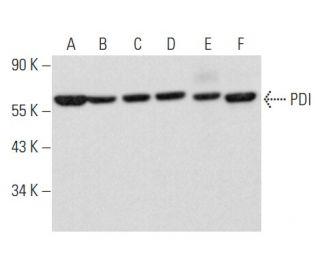
PDI Antibody (A-1) is a mouse monoclonal IgG1 antibody that detects protein disulfide isomerase (PDI) in mouse, rat, and human samples through various applications including western blotting (WB), immunoprecipitation (IP), immunofluorescence (IF), immunohistochemistry, and enzyme-linked immunosorbent assay (ELISA). Anti-PDI antibody (A-1) recognizes PDI, a crucial protein that plays a significant role in the folding and maturation of newly synthesized proteins within the endoplasmic reticulum (ER). PDI (A-1) antibody targets a protein essential for maintaining cellular homeostasis, as PDI catalyzes thiol-disulfide exchange reactions, which are vital for the proper formation of disulfide bonds in proteins. PDI (A-1) monoclonal antibody identifies a molecular chaperone that not only aids in the correct folding of proteins but also prevents the aggregation of misfolded proteins, thereby contributing to cellular stress responses. PDI monoclonal antibody (A-1) detects a protein involved in critical interactions with other proteins, such as immunoglobulin heavy chain binding protein (BiP), enhancing PDI′s role in protein processing and quality control within the ER. PDI′s localization in the ER is particularly important, as PDI assists in protein folding as they are synthesized, ensuring proteins reach their functional conformations efficiently. With PDI′s multifaceted roles in protein maturation and cellular signaling, anti-PDI antibody (A-1) serves as an invaluable tool for researchers studying protein dynamics and cellular function.
Ordering Information
| Product Name | Catalog # | UNIT | Price | Qty | FAVORITES | |
PDI Antibody (A-1) | sc-376370 | 200 µg/ml | $322.00 | |||
PDI Antibody (A-1): m-IgG Fc BP-HRP Bundle | sc-540555 | 200 µg Ab; 10 µg BP | $361.00 | |||
PDI Antibody (A-1): m-IgG1 BP-HRP Bundle | sc-542203 | 200 µg Ab; 20 µg BP | $361.00 | |||
PDI (A-1) Neutralizing Peptide | sc-376370 P | 100 µg/0.5 ml | $69.00 |