")
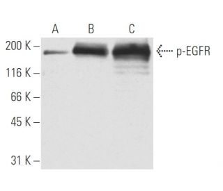
: sc-57544. Western Blot Analyse der EGFR-Phosphorylierung in unbehandelten (A), PMA-stimulierten (B) und Pervanadat-behandelten (C) OVCAR-5-Vollzelllysaten.")
p-EGFR Antikörper (9H2): sc-57545
- p-EGFR Antikörper 9H2 ist ein monoklonales IgG1 aus der Maus, verwendet in 12 wissenschaftlichen Veröffentlichungen, in einer Menge von 50 µg/0.5 ml
- gegen ein EFGR-Phosphopeptid mit Ursprung human
- Empfohlen für die Detektion von Tyr 1173 phosphorylated EGFR aus der Spezies mouse, rat, human und canine per WB, IP und IF; nicht kreuzreaktiv mit dem non-phosphorylated EGFR nor with unrelated Tyrosine-phosphorylated proteins
- Als Direktkonjugat zur Detektion von p-EGFR wird EGFR (A-10): sc-373746 angeboten; Primärantikörper konjugiert mit AC, HRP, FITC, PE, Alexa Fluor® 488, 594, 647, 680 und 790.
- m-IgG Fc BP-HRP und m-IgG1 BP-HRP sind die bevorzugten sekundären Nachweisreagenzien für p-EGFR Antikörper (9H2) for WB applications. Diese Reagenzien werden jetzt in Bündeln mit p-EGFR Antikörper (9H2) angeboten(siehe Bestellinformationen unten).

Direktverknüpfungen
Siehe auch...
Der p-EGFR-Antikörper (9H2) ist ein monoklonaler IgG1-Antikörper der Maus, der den phosphorylierten EGFR Tyr 1173 in Proben von Mäusen, Ratten, Menschen und Hunden durch Anwendungen wie Western Blotting (WB), Immunopräzipitation (IP) und Immunfluoreszenz (IF) nachweist. Der p-EGFR-monoklonale Antikörper (9H2) ist in nicht konjugierter Form erhältlich, was vielseitige Versuchsaufbauten ermöglicht. Der epidermale Wachstumsfaktor-Rezeptor (EGFR) spielt eine entscheidende Rolle bei der Signalübertragung in der Zelle und vermittelt zelluläre Reaktionen auf Wachstumsfaktoren, die für Prozesse wie Zellproliferation, Differenzierung und Überleben von entscheidender Bedeutung sind. Der hauptsächlich auf der Zelloberfläche befindliche EGFR wird durch die Bindung an Liganden wie EGF oder TGFα aktiviert, was eine Kaskade intrazellulärer Signalwege auslöst, die für die normale Zellfunktion und -entwicklung unerlässlich sind. Die Phosphorylierung spezifischer Tyrosinreste, insbesondere Tyr 1173, ist für die Aktivierung des EGFR und die anschließende Signalübertragung von entscheidender Bedeutung, da die Phosphorylierung die Rekrutierung nachgeschalteter Signalproteine erleichtert, darunter die Phosphotyrosin-Bindungsdomäne von GRB2. Diese Interaktion ist für die Aktivierung des Ras-Signalwegs von entscheidender Bedeutung, der für das Zellwachstum und die Zellteilung von wesentlicher Bedeutung ist. Eine Fehlregulation der EGFR-Signalübertragung ist häufig an verschiedenen Krebsarten beteiligt, sodass der p-EGFR (9H2)-Antikörper ein wertvolles Instrument für die Erforschung der Krebsbiologie und therapeutischer Interventionen darstellt.
Bestellinformation
| Produkt | Katalog # | EINHEIT | Preis | ANZAHL | Favoriten | |
p-EGFR Antikörper (9H2) | sc-57545 | 50 µg/0.5 ml | $322.00 | |||
p-EGFR (9H2): m-IgG Fc BP-HRP Bundle | sc-539060 | 50 µg Ab; 10 µg BP | $361.00 | |||
p-EGFR (9H2): m-IgG1 BP-HRP Bundle | sc-541235 | 50 µg Ab; 20 µg BP | $361.00 |