")
p-CD3-ζ Antibody (C415.9A): sc-9975
- p-CD3-ζ Antibody (C415.9A) is a mouse monoclonal IgG1 κ p-CD3-ζ antibody, cited in 17 publications, provided at 200 µg/ml
- raised against amino acids 52-163 mapping within the cytoplasmic domain of CD3-ζ precursor of human origin
- p-CD3-zeta Antibody (C415.9A) is recommended for detection of phosphorylated CD3-ζ of human origin by WB, IP, FCM and ELISA
- Anti-p-CD3-zeta Antibody (C415.9A) is available conjugated to agarose for IP; HRP for WB, IHC(P) and ELISA; and to either phycoerythrin or FITC for IF, IHC(P) and FCM
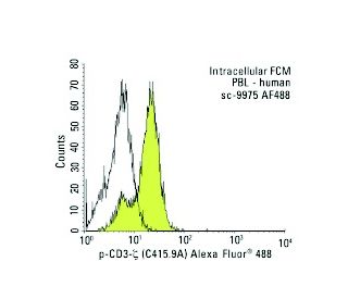
- also available conjugated to Alexa Fluor® 488, Alexa Fluor® 546, Alexa Fluor® 594 or Alexa Fluor® 647 for WB (RGB), IF, IHC(P) and FCM, and for use with RGB fluorescent imaging systems, such as iBright™ FL1000, FluorChem™, Typhoon, Azure and other comparable systems
- also available conjugated to Alexa Fluor® 680 or Alexa Fluor® 790 for WB (NIR), IF and FCM; for use with Near-Infrared (NIR) detection systems, such as LI-COR®Odyssey®, iBright™ FL1000, FluorChem™, Typhoon, Azure and other comparable systems
- m-IgG Fc BP-HRP, m-IgG1 BP-HRP and m-IgGκ BP-HRP are the preferred secondary detection reagents for p-CD3-ζ Antibody (C415.9A) for WB applications. These reagents are now offered in bundles with p-CD3-ζ Antibody (C415.9A) (see ordering information below).

QUICK LINKS
p-CD3-ζ Antibody (C415.9A) is a mouse monoclonal IgG1 kappa light chain antibody that detects p-CD3-zeta protein of human origin by western blotting (WB), immunoprecipitation (IP), flow cytometry (FCM), and enzyme-linked immunosorbent assay (ELISA). anti-p-CD3-ζ antibody (C415.9A) is available in both non-conjugated and various conjugated forms, including agarose, horseradish peroxidase (HRP), phycoerythrin (PE), fluorescein isothiocyanate (FITC), and multiple Alexa Fluor® conjugates. p-CD3-zeta protein plays a crucial role in T cell activation as a component of the T cell receptor (TCR) complex, which is essential for the immune response. p-CD3-zeta is located in the plasma membrane of T cells, where p-CD3-zeta forms part of a multisubunit complex that transmits signals upon recognition of foreign antigens. This signaling cascade is vital for the activation of T cells, leading to their proliferation and differentiation into effector cells that can target and eliminate pathogens. The zeta chain contains three immunoreceptor tyrosine-based activation motifs (ITAMs), which are critical for recruiting and activating downstream signaling molecules such as ZAP-70 and Syk. Phosphorylation of these ITAMs is a key step in the signaling process, allowing for the amplification of the immune response. Understanding p-CD3-zeta function and importance is essential for developing therapeutic strategies aimed at modulating T cell activity in various diseases, including cancer and autoimmune disorders.
Alexa Fluor® is a trademark of Molecular Probes Inc., OR., USA
LI-COR® and Odyssey® are registered trademarks of LI-COR Biosciences
p-CD3-ζ Antibody (C415.9A) References:
- Structure, assembly and intracellular transport of the T cell receptor for antigen. | Exley, M., et al. 1991. Semin Immunol. 3: 283-97. PMID: 1686832
- Signal transduction by the T cell antigen receptor. | Weiss, A., et al. 1991. Semin Immunol. 3: 313-24. PMID: 1839225
- Imaging T-cell receptor activation reveals accumulation of tyrosine-phosphorylated CD3ζ in the endosomal compartment. | Yudushkin, IA. and Vale, RD. 2010. Proc Natl Acad Sci U S A. 107: 22128-33. PMID: 21135224
- TCR crosslinking promotes Crk adaptor protein binding to tyrosine-phosphorylated CD3ζ chain. | Dong, G., et al. 2017. Biochem Biophys Res Commun. 488: 541-546. PMID: 28526413
- CD28 Costimulation Augments CAR Signaling in NK Cells via the LCK/CD3ζ/ZAP70 Signaling Axis. | Acharya, S., et al. 2024. Cancer Discov. 14: 1879-1900. PMID: 38900051
- Specific interaction of the CD45 protein-tyrosine phosphatase with tyrosine-phosphorylated CD3 zeta chain. | Furukawa, T., et al. 1994. Proc Natl Acad Sci U S A. 91: 10928-32. PMID: 7526385
- Signal transduction. Zapping tandem SH2 domains. | Weiss, A. 1995. Nature. 377: 17-8. PMID: 7659151
- Molecular basis for interaction of the protein tyrosine kinase ZAP-70 with the T-cell receptor. | Hatada, MH., et al. 1995. Nature. 377: 32-8. PMID: 7659156
- Binding of ZAP-70 to phosphorylated T-cell receptor zeta and eta enhances its autophosphorylation and generates specific binding sites for SH2 domain-containing proteins. | Neumeister, EN., et al. 1995. Mol Cell Biol. 15: 3171-8. PMID: 7760813
- Different cytoplasmic structure of the CD3 zeta family dimer modulates the activation signal and function of T cells. | Aoe, T., et al. 1994. Int Immunol. 6: 1671-9. PMID: 7865460
- The role of protein tyrosine kinases and protein tyrosine phosphatases in T cell antigen receptor signal transduction. | Chan, AC., et al. 1994. Annu Rev Immunol. 12: 555-92. PMID: 8011291
- Targeted disruption of the CD3 eta locus causes high lethality in mice: modulation of Oct-1 transcription on the opposite strand. | Ohno, H., et al. 1994. EMBO J. 13: 1157-65. PMID: 8131747
Ordering Information
| Product Name | Catalog # | UNIT | Price | Qty | FAVORITES | |
p-CD3-ζ Antibody (C415.9A) | sc-9975 | 200 µg/ml | $322.00 | |||
p-CD3-ζ Antibody (C415.9A): m-IgG Fc BP-HRP Bundle | sc-528243 | 200 µg Ab; 10 µg BP | $361.00 | |||
p-CD3-ζ Antibody (C415.9A): m-IgGκ BP-HRP Bundle | sc-520570 | 200 µg Ab, 40 µg BP | $361.00 | |||
p-CD3-ζ Antibody (C415.9A): m-IgG1 BP-HRP Bundle | sc-542833 | 200 µg Ab; 20 µg BP | $361.00 | |||
p-CD3-ζ Antibody (C415.9A) AC | sc-9975 AC | 500 µg/ml, 25% agarose | $424.00 | |||
p-CD3-ζ Antibody (C415.9A) HRP | sc-9975 HRP | 200 µg/ml | $322.00 | |||
p-CD3-ζ Antibody (C415.9A) FITC | sc-9975 FITC | 200 µg/ml | $336.00 | |||
p-CD3-ζ Antibody (C415.9A) PE | sc-9975 PE | 200 µg/ml | $349.00 | |||
p-CD3-ζ Antibody (C415.9A) Alexa Fluor® 488 | sc-9975 AF488 | 200 µg/ml | $364.00 | |||
p-CD3-ζ Antibody (C415.9A) Alexa Fluor® 546 | sc-9975 AF546 | 200 µg/ml | $364.00 | |||
p-CD3-ζ Antibody (C415.9A) Alexa Fluor® 594 | sc-9975 AF594 | 200 µg/ml | $364.00 | |||
p-CD3-ζ Antibody (C415.9A) Alexa Fluor® 647 | sc-9975 AF647 | 200 µg/ml | $364.00 | |||
p-CD3-ζ Antibody (C415.9A) Alexa Fluor® 680 | sc-9975 AF680 | 200 µg/ml | $364.00 | |||
p-CD3-ζ Antibody (C415.9A) Alexa Fluor® 790 | sc-9975 AF790 | 200 µg/ml | $364.00 |