")
p-Akt1/2/3 Antibody (B-5): sc-271966
- p-Akt1/2/3 Antibody (B-5) is a mouse monoclonal IgG2b κ, cited in 114 publications, provided at 200 µg/ml
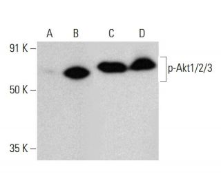
- p-Akt1/2/3 Antibody (B-5) is recommended for detection of Thr 308 phosphorylated Akt1 and correspondingly Thr 309 phosphorylated Akt2 and correspondingly Thr 305 phosphorylated Akt3 of mouse, rat, human and origin by WB, IP, IF, IHC(P) and ELISA; also reactive with additional species, including equine and avian
- Anti-p-Akt1/2/3 Antibody (B-5) is available conjugated to agarose for IP; HRP for WB, IHC(P) and ELISA; and to either phycoerythrin or FITC for IF, IHC(P) and FCM
- also available conjugated to Alexa Fluor® 488, Alexa Fluor® 546, Alexa Fluor® 594 or Alexa Fluor® 647 for WB (RGB), IF, IHC(P) and FCM, and for use with RGB fluorescent imaging systems, such as iBright™ FL1000, FluorChem™, Typhoon, Azure and other comparable systems
- also available conjugated to Alexa Fluor® 680 or Alexa Fluor® 790 for WB (NIR), IF and FCM; for use with Near-Infrared (NIR) detection systems, such as LI-COR®Odyssey®, iBright™ FL1000, FluorChem™, Typhoon, Azure and other comparable systems
- m-IgG2b BP-HRP is the preferred secondary detection reagent for p-Akt1/2/3 Antibody (B-5) for WB and IHC(P) applications. This reagent is now offered in a bundle with p-Akt1/2/3 Antibody (B-5) (see ordering information below).

p-Akt1/2/3 Antibody (B-5) is a mouse monoclonal IgG2b kappa light chain antibody that detects p-Akt1/2/3, specifically Thr 308 phosphorylated Akt1, Thr 309 phosphorylated Akt2, and Thr 305 phosphorylated Akt3, in mouse, rat, and human samples through applications such as western blotting (WB), immunoprecipitation (IP), immunofluorescence (IF), immunohistochemistry, and enzyme-linked immunosorbent assay (ELISA). p-Akt1/2/3 (B-5) antibody is available in both non-conjugated and various conjugated forms, including agarose, horseradish peroxidase (HRP), phycoerythrin (PE), fluorescein isothiocyanate (FITC), and multiple Alexa Fluor® conjugates. The Akt family of serine/threonine kinases, which includes Akt1, Akt2, and Akt3, plays a crucial role in cellular signaling pathways that regulate metabolism, growth, and survival. Notably, Akt proteins are activated by insulin and insulin-like growth factor-1 (IGF-1), leading to phosphorylation at specific threonine and serine residues, which is essential for their function in promoting glucose uptake and glycogen synthesis. The pleckstrin homology domain present in all Akt family members is vital for their membrane localization and subsequent activation by phosphatidylinositol 3-kinase (PI3K) signaling. Akt3, while also responsive to insulin, has unique regulatory mechanisms that differentiate its activation from that of Akt1 and Akt2, particularly in the context of protein kinase C signaling. Understanding the intricate roles of these proteins in various biological processes, including cellular proliferation and apoptosis, underscores the importance of anti-p-Akt1/2/3 antibody (B-5) in research focused on metabolic diseases, cancer, and other conditions where Akt signaling is disrupted.
Alexa Fluor® is a trademark of Molecular Probes Inc., OR., USA
LI-COR® and Odyssey® are registered trademarks of LI-COR Biosciences
p-Akt1/2/3 Antibody (B-5) References:
- Cytoplasmic Skp2 expression is associated with p-Akt1 and predicts poor prognosis in human breast carcinomas. | Liu, J., et al. 2012. PLoS One. 7: e52675. PMID: 23300741
- Discovery of 4-(Piperazin-1-yl)-7H-pyrrolo[2,3-d]pyrimidine Derivatives as Akt Inhibitors. | Liu, Y., et al. 2016. Arch Pharm (Weinheim). 349: 356-62. PMID: 26991997
- Slug inhibits the proliferation and tumor formation of human cervical cancer cells by up-regulating the p21/p27 proteins and down-regulating the activity of the Wnt/β-catenin signaling pathway via the trans-suppression Akt1/p-Akt1 expression. | Cui, N., et al. 2016. Oncotarget. 7: 26152-67. PMID: 27036045
- Quercetin Inhibits the Migration and Invasion of HCCLM3 Cells by Suppressing the Expression of p-Akt1, Matrix Metalloproteinase (MMP) MMP-2, and MMP-9. | Lu, J., et al. 2018. Med Sci Monit. 24: 2583-2589. PMID: 29701200
- P-AKT2/SPK1 (P-SPK1) and P-MEK/P-ERK cell signaling pathways are involved in LPS-induced macrophage migration. | Lei, Y., et al. 2019. Am J Transl Res. 11: 2725-2741. PMID: 31217849
- Expression of Akt1 and p-Akt1 in peripheral T cell subsets of multiple sclerosis patients. | Oktelik, FB., et al. 2021. Acta Neurol Belg. 121: 1777-1782. PMID: 33034831
- Hexokinases 2 promoted cell motility and distant metastasis by elevating fibronectin through Akt1/p-Akt1 in cervical cancer cells. | Chen, Q., et al. 2021. Cancer Cell Int. 21: 600. PMID: 34758823
- Hexokinase 2 promoted cell motility and proliferation by activating Akt1/p-Akt1 in human ovarian cancer cells. | Tian, X., et al. 2022. J Ovarian Res. 15: 92. PMID: 35953860
- The Significance of p-AKT1 as a Prognostic Marker and Therapeutic Target in Patients With Hormone Receptor-Positive and Human Epidermal Growth Factor Receptor-2-Positive Early Breast Cancer. | Kim, JY., et al. 2022. J Breast Cancer. 25: 387-403. PMID: 36314765
- IL-21/23 axis modulates inflammatory cytokines and RANKL expression in RA CD4+ T cells via p-Akt1 signaling. | Bhattacharya, G., et al. 2023. Front Immunol. 14: 1235514. PMID: 37809066
Ordering Information
| Product Name | Catalog # | UNIT | Price | Qty | FAVORITES | |
p-Akt1/2/3 Antibody (B-5) | sc-271966 | 200 µg/ml | $322.00 | |||
p-Akt1/2/3 Antibody (B-5): m-IgG2b BP-HRP Bundle | sc-548711 | 200 µg Ab; 10 µg BP | $361.00 | |||
p-Akt1/2/3 Antibody (B-5) AC | sc-271966 AC | 500 µg/ml, 25% agarose | $424.00 | |||
p-Akt1/2/3 Antibody (B-5) HRP | sc-271966 HRP | 200 µg/ml | $322.00 | |||
p-Akt1/2/3 Antibody (B-5) FITC | sc-271966 FITC | 200 µg/ml | $336.00 | |||
p-Akt1/2/3 Antibody (B-5) PE | sc-271966 PE | 200 µg/ml | $349.00 | |||
p-Akt1/2/3 Antibody (B-5) Alexa Fluor® 488 | sc-271966 AF488 | 200 µg/ml | $364.00 | |||
p-Akt1/2/3 Antibody (B-5) Alexa Fluor® 546 | sc-271966 AF546 | 200 µg/ml | $364.00 | |||
p-Akt1/2/3 Antibody (B-5) Alexa Fluor® 594 | sc-271966 AF594 | 200 µg/ml | $364.00 | |||
p-Akt1/2/3 Antibody (B-5) Alexa Fluor® 647 | sc-271966 AF647 | 200 µg/ml | $364.00 | |||
p-Akt1/2/3 Antibody (B-5) Alexa Fluor® 680 | sc-271966 AF680 | 200 µg/ml | $364.00 | |||
p-Akt1/2/3 Antibody (B-5) Alexa Fluor® 790 | sc-271966 AF790 | 200 µg/ml | $364.00 | |||
p-Akt1/2/3 (B-5) Neutralizing Peptide | sc-271966 P | 100 µg/0.5 ml | $69.00 |