")
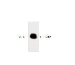
: sc-136140. Western blot analysis of Nir2 expression in rat cerebrum tissue extract.")
: sc-136140. Western blot analysis of Nir2 expression in LADMAC whole cell lysate (A) and rat cerebellum tissue extract (B). Detection reagent used: m-IgGκ BP-HRP: sc-516102.")
Nir2 Antibody (8): sc-136140
- Nir2 Antibody (8) is a mouse monoclonal IgG1 κ Nir2 antibody provided at 200 µg/ml
- raised against amino acids 235-332 of Nir2 of mouse origin
- Nir2 Antibody (8) is recommended for detection of Nir2 of mouse and rat origin by WB and IP
- m-IgGκ BP-HRP is the preferred secondary detection reagent for Nir2 Antibody (8) for WB applications. This reagent is now offered in a bundle with Nir2 Antibody (8) (see ordering information below). For additional m-IgGκ BP conjugates see our complete list of Mouse IgG Binding Proteins.

QUICK LINKS
Nir2 Antibody (8) is a mouse monoclonal IgG1 kappa light chain antibody that detects Nir2 protein of mouse and rat origin by western blotting (WB) and immunoprecipitation (IP). Anti-Nir2 antibody (8) is available as non-conjugated format. Nir proteins, including Nir2, are human homologues of Drosophila retinal degeneration B (rdgB) and have been implicated as candidate genes for inherited retinal degeneration diseases. Nir2 is primarily localized in the Golgi apparatus during interphase, playing a crucial role in processing and trafficking proteins within cells. During cytokinesis, Nir2 is recruited to the cleavage furrow and midbody, highlighting its importance in cell division. Structurally, Nir2 features an amino-terminal phosphatidylinositol-transfer protein (PITP)-like domain, an acidic calcium-binding domain, six putative transmembrane domains, and a conserved carboxyl-terminal domain, suggesting involvement in calcium and phosphoinositide metabolism downstream of G-protein-coupled receptors. These unique localization and structural characteristics underscore Nir2′s essential function in cellular processes, making Nir2 a valuable target for research in retinal health and disease.
Alexa Fluor® is a trademark of Molecular Probes Inc., OR., USA
LI-COR® and Odyssey® are registered trademarks of LI-COR Biosciences
Nir2 Antibody (8) References:
- Cloning and characterization of a novel human phosphatidylinositol transfer protein, rdgBbeta. | Fullwood, Y., et al. 1999. J Biol Chem. 274: 31553-8. PMID: 10531358
- Nir2, a novel regulator of cell morphogenesis. | Tian, D., et al. 2002. Mol Cell Biol. 22: 2650-62. PMID: 11909959
- Cellular and developmental distribution of human homologues of the Drosophilia rdgB protein in the rat retina. | Tian, D. and Lev, S. 2002. Invest Ophthalmol Vis Sci. 43: 1946-53. PMID: 12037004
- Nir2, a human homolog of Drosophila melanogaster retinal degeneration B protein, is essential for cytokinesis. | Litvak, V., et al. 2002. Mol Cell Biol. 22: 5064-75. PMID: 12077336
- Targeting of Nir2 to lipid droplets is regulated by a specific threonine residue within its PI-transfer domain. | Litvak, V., et al. 2002. Curr Biol. 12: 1513-8. PMID: 12225667
Ordering Information
| Product Name | Catalog # | UNIT | Price | Qty | FAVORITES | |
Nir2 Antibody (8) | sc-136140 | 200 µg/ml | $322.00 | |||
Nir2 Antibody (8): m-IgGκ BP-HRP Bundle | sc-521331 | 200 µg Ab, 40 µg BP | $361.00 |