")
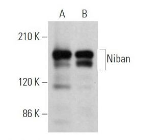
Niban Antibody (F-10): sc-374636
- Niban Antibody (F-10) is a mouse monoclonal IgG1 κ Niban antibody provided at 200 µg/ml
- raised against amino acids 1-240 mapping at the N-terminus of Niban of human origin
- Niban Antibody (F-10) is recommended for detection of Niban of human origin by WB, IP, IF and ELISA
- Anti-Niban Antibody (F-10) is available conjugated to agarose for IP; HRP for WB, IHC(P) and ELISA; and to either phycoerythrin or FITC for IF, IHC(P) and FCM
- also available conjugated to Alexa Fluor® 488, Alexa Fluor® 546, Alexa Fluor® 594 or Alexa Fluor® 647 for WB (RGB), IF, IHC(P) and FCM, and for use with RGB fluorescent imaging systems, such as iBright™ FL1000, FluorChem™, Typhoon, Azure and other comparable systems
- also available conjugated to Alexa Fluor® 680 or Alexa Fluor® 790 for WB (NIR), IF and FCM; for use with Near-Infrared (NIR) detection systems, such as LI-COR®Odyssey®, iBright™ FL1000, FluorChem™, Typhoon, Azure and other comparable systems
- m-IgG Fc BP-HRP and m-IgG1 BP-HRP are the preferred secondary detection reagents for Niban Antibody (F-10) for WB applications. These reagents are now offered in bundles with Niban Antibody (F-10) (see ordering information below).

QUICK LINKS
SEE ALSO...
Niban Antibody (F-10) is a mouse monoclonal IgG1 kappa light chain antibody that detects Niban protein of human origin by western blotting (WB), immunoprecipitation (IP), immunofluorescence (IF), and enzyme-linked immunosorbent assay (ELISA). Niban (F-10) antibody is available in both non-conjugated and various conjugated forms, including agarose, horseradish peroxidase (HRP), phycoerythrin (PE), fluorescein isothiocyanate (FITC), and multiple Alexa Fluor® conjugates. Known as FAM129A and Cell growth-inhibiting gene 39 protein, Niban is a 928 amino acid cytoplasmic protein that plays a crucial role in regulating phosphorylation of key proteins involved in translation regulation, such as eIF2α, 4E-BP1, and p70 S6 kinase α. Niban′s ability to modulate these phosphorylation events is vital for maintaining cellular homeostasis and preventing uncontrolled cell growth, which is particularly important in cancer. Niban has been implicated as a tumor marker for various cancers, including renal carcinoma, thyroid cancer, and head and neck squamous cell carcinoma. Notably, endoplasmic reticular stress in Niban knockout mice results in upregulation of eIF2α and decreased phosphorylation of p70 S6 kinase α and 4E-BP1, leading to increased apoptosis. This highlights Niban′s significant role in modulating cell death signaling pathways through regulatory effects on translation, making Niban a critical protein for understanding cancer biology and potential therapeutic targets.
Alexa Fluor® is a trademark of Molecular Probes Inc., OR., USA
LI-COR® and Odyssey® are registered trademarks of LI-COR Biosciences
Niban Antibody (F-10) References:
- A novel gene 'Niban' upregulated in renal carcinogenesis: cloning by the cDNA-amplified fragment length polymorphism approach. | Majima, S., et al. 2000. Jpn J Cancer Res. 91: 869-74. PMID: 11011112
- Niban gene is commonly expressed in the renal tumors: a new candidate marker for renal carcinogenesis. | Adachi, H., et al. 2004. Oncogene. 23: 3495-500. PMID: 14990989
- Multistep renal carcinogenesis in the Eker (Tsc 2 gene mutant) rat model. | Hino, O. 2004. Curr Mol Med. 4: 807-11. PMID: 15579027
- Hepatic angiomyolipoma and hepatic stellate cells share a similar gene expression profile. | Kannangai, R., et al. 2005. Hum Pathol. 36: 341-7. PMID: 15891994
- A novel tumor marker, Niban, is expressed in subsets of thyroid tumors and Hashimoto's thyroiditis. | Matsumoto, F., et al. 2006. Hum Pathol. 37: 1592-600. PMID: 16949643
- The endoplasmic reticulum stress-inducible protein Niban regulates eIF2alpha and S6K1/4E-BP1 phosphorylation. | Sun, GD., et al. 2007. Biochem Biophys Res Commun. 360: 181-7. PMID: 17588536
- Frequent expression of Niban in head and neck squamous cell carcinoma and squamous dysplasia. | Ito, S., et al. 2010. Head Neck. 32: 96-103. PMID: 19536772
Ordering Information
| Product Name | Catalog # | UNIT | Price | Qty | FAVORITES | |
Niban Antibody (F-10) | sc-374636 | 200 µg/ml | $322.00 | |||
Niban Antibody (F-10): m-IgG Fc BP-HRP Bundle | sc-527495 | 200 µg Ab; 10 µg BP | $361.00 | |||
Niban Antibody (F-10): m-IgG1 BP-HRP Bundle | sc-532868 | 200 µg Ab; 20 µg BP | $361.00 | |||
Niban Antibody (F-10) AC | sc-374636 AC | 500 µg/ml, 25% agarose | $424.00 | |||
Niban Antibody (F-10) HRP | sc-374636 HRP | 200 µg/ml | $322.00 | |||
Niban Antibody (F-10) FITC | sc-374636 FITC | 200 µg/ml | $336.00 | |||
Niban Antibody (F-10) PE | sc-374636 PE | 200 µg/ml | $349.00 | |||
Niban Antibody (F-10) Alexa Fluor® 488 | sc-374636 AF488 | 200 µg/ml | $364.00 | |||
Niban Antibody (F-10) Alexa Fluor® 546 | sc-374636 AF546 | 200 µg/ml | $364.00 | |||
Niban Antibody (F-10) Alexa Fluor® 594 | sc-374636 AF594 | 200 µg/ml | $364.00 | |||
Niban Antibody (F-10) Alexa Fluor® 647 | sc-374636 AF647 | 200 µg/ml | $364.00 | |||
Niban Antibody (F-10) Alexa Fluor® 680 | sc-374636 AF680 | 200 µg/ml | $364.00 | |||
Niban Antibody (F-10) Alexa Fluor® 790 | sc-374636 AF790 | 200 µg/ml | $364.00 |