")
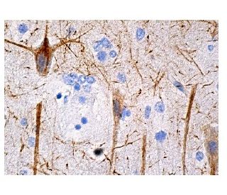
: sc-133165. Tinción de inmunoperoxidasa de tejido cerebral humano fijado con formalina e incluido en parafina que muestra tinción citoplasmática de células neuronales.")
: sc-133165. Análisis por Western blot de la expresión de NF-H en lisados de células enteras de 293T no transfectadas: sc-117752 (A), 293T transfectadas con NF-H humano: sc-111457 (B) y extracto de tejido de cerebro de rata (C).")
: sc-133165. Análisis por Western blot de la expresión de NF-H en extracto de tejido cerebral humano.")
NF-H Anticuerpo (A-12): sc-133165
- NF-H Anticuerpo (A-12) es un monoclonal de ratón IgG1 κ, ver las 2 publicaciones, proporcionado como 200 µg/ml
- planteada frente a los aminoácidos 1-100 mapeados en el N-terminus de NF-H de human origen
- recomendado para detectarNF-H de mouse, rat y human origen, mediante WB, IP, IF, IHC(P) y ELISA
- m-IgG Fc BP-HRP y m-IgG1 BP-HRP son los reactivos de detección secundarios preferidos para NF-H Anticuerpo (A-12) for WB and IHC(P) applications. Estos reactivos se ofrecen ahora en paquetes con NF-H Anticuerpo (A-12)(véase la información de pedido más abajo).

Neurofilament-H (NF-H), para el polipéptido pesado de neurofilamento, un miembro de la familia de filamentos intermedios, es un componente importante de los citoesqueletos neuronales. Los neurofilamentos son estructuras dinámicas; contienen sitios de fosforilación para un gran número de proteínas quinasas, incluyendo la proteína quinasa A, la proteína quinasa C, la quinasa dependiente de ciclina 5, la quinasa regulada por señales extracelulares, la quinasa de la glicógeno sintasa-3 y la quinasa de la proteína activada por estrés gamma. Además de su papel en el control del calibre del axón, los neurofilamentos pueden afectar a otros elementos del citoesqueleto, como los microtúbulos y los filamentos de actina. Los cambios en la fosforilación o el metabolismo de los neurofilamentos se observan con frecuencia en enfermedades neurodegenerativas, incluyendo la esclerosis lateral amiotrófica (ELA), la enfermedad de Parkinson y la enfermedad de Alzheimer.
Alexa Fluor® es una marca registrada de Molecular Probes Inc., OR., USA
REIVEW LI-COR® y Odyssey® son marcas registradas de LI-COR Biosciences.
NF-H Anticuerpo (A-12) Referencias:
- Las subunidades extra de neurofilamento NF-L rescatan la enfermedad de la neurona motora causada por la sobreexpresión del gen NF-H humano en ratones. | Meier, J., et al. 1999. J Neuropathol Exp Neurol. 58: 1099-110. PMID: 10515233
- La sobreexpresión de las subunidades NF-L y NF-H de los neurofilamentos prolonga la supervivencia de un modelo de ratón para la esclerosis lateral amiotrófica. | Kong, J. and Xu, Z. 2000. Neurosci Lett. 281: 72-4. PMID: 10686419
- El dominio de la cola C-terminal de la proteína neurofilamento-H (NF-H) forma los puentes cruzados y regula la formación de haces de neurofilamentos. | Chen, J., et al. 2000. J Cell Sci. 113 Pt 21: 3861-9. PMID: 11034913
- La forma axonal fosforilada de la subunidad de neurofilamentos NF-H (pNF-H) como biomarcador sanguíneo de la lesión cerebral traumática. | Anderson, KJ., et al. 2008. J Neurotrauma. 25: 1079-85. PMID: 18729720
- La subunidad NF-H fosforilada de los neurofilamentos como biomarcador para evaluar la gravedad de los pacientes con lesiones de la médula espinal, un estudio piloto. | Hayakawa, K., et al. 2012. Spinal Cord. 50: 493-6. PMID: 22270191
- El neurofilamento de alto peso molecular (NF-H) transfectado en ratas se coensambla con vimentina en una forma predominantemente no fosforilada. | Chin, SS. and Liem, RK. 1990. J Neurosci. 10: 3714-26. PMID: 2230956
- Fosforilación de la proteína neurofilamento de alto peso molecular (NF-H) por Cdk5 y p35. | Sun, D., et al. 1996. J Biol Chem. 271: 14245-51. PMID: 8662984
- Propiedades de ensamblaje de proteínas de neurofilamento NF-H truncadas en los extremos amino y carboxilo con NF-L y NF-M en presencia y ausencia de vimentina. | Sun, D., et al. 1997. J Neurochem. 68: 917-26. PMID: 9048736
- Papeles antagónicos de las subunidades de neurofilamentos NF-H y NF-M frente a NF-L en la conformación de la arborización dendrítica en las neuronas motoras espinales. | Kong, J., et al. 1998. J Cell Biol. 140: 1167-76. PMID: 9490729
- Las proteínas quinasas activadas por mitógenos (Erk1,2) fosforilan las repeticiones Lys-Ser-Pro (KSP) en las proteínas neurofilamentosas NF-H y NF-M. | Veeranna,., et al. 1998. J Neurosci. 18: 4008-21. PMID: 9592082
Información sobre pedidos
| Nombre del producto | Número de catálogo | UNIDAD | Precio | CANTIDAD | Favoritos | |
NF-H Anticuerpo (A-12) | sc-133165 | 200 µg/ml | $322.00 | |||
Paquete de NF-H (A-12): m-IgG Fc BP-HRP | sc-539859 | 200 µg Ab; 10 µg BP | $361.00 | |||
Paquete de NF-H (A-12): m-IgG1 BP-HRP | sc-541751 | 200 µg Ab; 20 µg BP | $361.00 |