")
MICAL1 Antibody (D-3): sc-515814
- MICAL1 Antibody (D-3) is a mouse monoclonal IgG2a κ MICAL1 antibody provided at 200 µg/ml
- specific for an epitope mapping between amino acids 768-792 within an internal region of MICAL1 of human origin
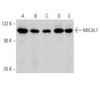
- MICAL1 Antibody (D-3) is recommended for detection of MICAL1 of mouse, rat and human origin by WB, IP, IF and ELISA
- Anti-MICAL1 Antibody (D-3) is available conjugated to agarose for IP; HRP for WB, IHC(P) and ELISA; and to either phycoerythrin or FITC for IF, IHC(P) and FCM
- also available conjugated to Alexa Fluor® 488, Alexa Fluor® 546, Alexa Fluor® 594 or Alexa Fluor® 647 for WB (RGB), IF, IHC(P) and FCM, and for use with RGB fluorescent imaging systems, such as iBright™ FL1000, FluorChem™, Typhoon, Azure and other comparable systems
- also available conjugated to Alexa Fluor® 680 or Alexa Fluor® 790 for WB (NIR), IF and FCM; for use with Near-Infrared (NIR) detection systems, such as LI-COR®Odyssey®, iBright™ FL1000, FluorChem™, Typhoon, Azure and other comparable systems
- m-IgG Fc BP-HRP, m-IgG2a BP-HRP and m-IgGκ BP-HRP are the preferred secondary detection reagents for MICAL1 Antibody (D-3) for WB applications. These reagents are now offered in bundles with MICAL1 Antibody (D-3) (see ordering information below).

QUICK LINKS
MICAL1 Antibody (D-3) is a mouse monoclonal IgG2a kappa light chain antibody that detects MICAL1 protein of mouse, rat, and human origin by western blotting (WB), immunoprecipitation (IP), immunofluorescence (IF), and enzyme-linked immunosorbent assay (ELISA). MICAL1 Antibody (D-3) is available in both non-conjugated and various conjugated forms, including agarose, horseradish peroxidase (HRP), phycoerythrin (PE), fluorescein isothiocyanate (FITC), and multiple Alexa Fluor® conjugates. MICAL1, or microtubule associated monooxygenase, calponin and LIM domain containing 1, is a 1,067 amino acid protein that plays a crucial role in cytoskeletal regulation, localizing to both the cytoplasm and the cytoskeleton. MICAL1 contains one LIM zinc-binding domain and one calponin-homology domain, which are essential for interactions with other proteins. MICAL1 is expressed in various tissues, including the kidney, thymus, spleen, lung, and testis, and interacts with several key proteins such as the SH3 domain of Cas-L, Rab 1B, plexin-A3, and Vimentin. These interactions are vital for regulating cytoskeletal dynamics and may contribute to processes like axonal repulsion, highlighting MICAL1′s importance in cellular architecture and signaling pathways. Notably, three isoforms of MICAL1 exist due to alternative splicing, which may further diversify functional roles in different cellular contexts.
Alexa Fluor® is a trademark of Molecular Probes Inc., OR., USA
LI-COR® and Odyssey® are registered trademarks of LI-COR Biosciences
MICAL1 Antibody (D-3) References:
- MICAL, a novel CasL interacting molecule, associates with vimentin. | Suzuki, T., et al. 2002. J Biol Chem. 277: 14933-41. PMID: 11827972
- MICALs, a family of conserved flavoprotein oxidoreductases, function in plexin-mediated axonal repulsion. | Terman, JR., et al. 2002. Cell. 109: 887-900. PMID: 12110185
- MICAL-1 isoforms, novel rab1 interacting proteins. | Weide, T., et al. 2003. Biochem Biophys Res Commun. 306: 79-86. PMID: 12788069
- The MICAL proteins and rab1: a possible link to the cytoskeleton? | Fischer, J., et al. 2005. Biochem Biophys Res Commun. 328: 415-23. PMID: 15694364
- Identification and characterization of Iporin as a novel interaction partner for rab1. | Bayer, M., et al. 2005. BMC Cell Biol. 6: 15. PMID: 15796781
- Investigation of the four cooperative unfolding units existing in the MICAL-1 CH domain. | Jin, X., et al. 2007. Biophys Chem. 129: 269-78. PMID: 17662518
- Release of MICAL autoinhibition by semaphorin-plexin signaling promotes interaction with collapsin response mediator protein. | Schmidt, EF., et al. 2008. J Neurosci. 28: 2287-97. PMID: 18305261
Ordering Information
| Product Name | Catalog # | UNIT | Price | Qty | FAVORITES | |
MICAL1 Antibody (D-3) | sc-515814 | 200 µg/ml | $322.00 | |||
MICAL1 Antibody (D-3): m-IgG Fc BP-HRP Bundle | sc-531522 | 200 µg Ab; 10 µg BP | $361.00 | |||
MICAL1 Antibody (D-3): m-IgGκ BP-HRP Bundle | sc-525233 | 200 µg Ab, 40 µg BP | $361.00 | |||
MICAL1 Antibody (D-3): m-IgG2a BP-HRP Bundle | sc-547810 | 200 µg Ab; 10 µg BP | $361.00 | |||
MICAL1 Antibody (D-3) AC | sc-515814 AC | 500 µg/ml, 25% agarose | $424.00 | |||
MICAL1 Antibody (D-3) HRP | sc-515814 HRP | 200 µg/ml | $322.00 | |||
MICAL1 Antibody (D-3) FITC | sc-515814 FITC | 200 µg/ml | $336.00 | |||
MICAL1 Antibody (D-3) PE | sc-515814 PE | 200 µg/ml | $349.00 | |||
MICAL1 Antibody (D-3) Alexa Fluor® 488 | sc-515814 AF488 | 200 µg/ml | $364.00 | |||
MICAL1 Antibody (D-3) Alexa Fluor® 546 | sc-515814 AF546 | 200 µg/ml | $364.00 | |||
MICAL1 Antibody (D-3) Alexa Fluor® 594 | sc-515814 AF594 | 200 µg/ml | $364.00 | |||
MICAL1 Antibody (D-3) Alexa Fluor® 647 | sc-515814 AF647 | 200 µg/ml | $364.00 | |||
MICAL1 Antibody (D-3) Alexa Fluor® 680 | sc-515814 AF680 | 200 µg/ml | $364.00 | |||
MICAL1 Antibody (D-3) Alexa Fluor® 790 | sc-515814 AF790 | 200 µg/ml | $364.00 |