")
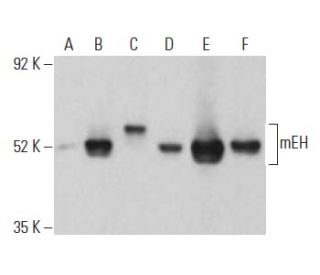
mEH Antibody (17): sc-135984
- mEH Antibody (17) is a mouse monoclonal IgG1 κ mEH antibody, cited in 8 publications, provided at 200 µg/ml
- raised against amino acids 13-125 of mEH of human origin
- recommended for detection of mEH of mouse, rat and human origin by WB, IP and IF
- m-IgGκ BP-HRP is the preferred secondary detection reagent for mEH Antibody (17) for WB applications. This reagent is now offered in a bundle with mEH Antibody (17) (see ordering information below). For additional m-IgGκ BP conjugates see our complete list of Mouse IgG Binding Proteins.

QUICK LINKS
SEE ALSO...
mEH Antibody (17) is a mouse monoclonal IgG1 kappa light chain antibody that detects mEH protein of mouse, rat, and human origin by western blotting (WB), immunoprecipitation (IP), and immunofluorescence (IF). anti-mEH antibody (17) is available as the non-conjugated form. mEH protein, also known as microsomal epoxide hydrolase, plays a crucial role in the detoxification of harmful compounds by catalyzing the hydrolysis of epoxides into less reactive and more water-soluble dihydrodiols, which can then be easily excreted from the body. This enzymatic process is vital for protecting cells from the potentially damaging effects of various endogenous and exogenous toxicants, including those derived from tobacco smoke. mEH protein is encoded by the EPHX1 gene, located on chromosome 1q42.12, and exhibits broad substrate specificity, allowing mEH protein to metabolize a wide range of substrates. Notably, polymorphisms in the EPHX1 gene have been associated with an increased risk of certain cancers, such as ovarian cancer and hepatocellular carcinoma, highlighting the importance of mEH in both metabolic processes and disease susceptibility.
Alexa Fluor® is a trademark of Molecular Probes Inc., OR., USA
LI-COR® and Odyssey® are registered trademarks of LI-COR Biosciences
mEH Antibody (17) References:
- Analysis of the EPHX1 113 polymorphism and GSTM1 homozygous null polymorphism and oral clefting associated with maternal smoking. | Hartsfield, JK., et al. 2001. Am J Med Genet. 102: 21-4. PMID: 11471167
- Inhibitors of soluble epoxide hydrolase attenuate vascular smooth muscle cell proliferation. | Davis, BB., et al. 2002. Proc Natl Acad Sci U S A. 99: 2222-7. PMID: 11842228
- Microsomal epoxide hydrolase polymorphism as a risk factor for ovarian cancer. | Lancaster, JM., et al. 1996. Mol Carcinog. 17: 160-2. PMID: 8944076
- The role of human glutathione transferases and epoxide hydrolases in the metabolism of xenobiotics. | Seidegård, J. and Ekström, G. 1997. Environ Health Perspect. 105 Suppl 4: 791-9. PMID: 9255563
Ordering Information
| Product Name | Catalog # | UNIT | Price | Qty | FAVORITES | |
mEH Antibody (17) | sc-135984 | 200 µg/ml | $322.00 | |||
mEH Antibody (17): m-IgGκ BP-HRP Bundle | sc-521315 | 200 µg Ab, 40 µg BP | $361.00 |