
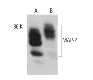
MAP-2 Antibody (C-2): sc-390543
- MAP-2 Antibody (C-2) is a mouse monoclonal IgG1 κ, cited in 5 publications, provided at 200 µg/ml
- specific for an epitope mapping between amino acids 1754-1777 of MAP-2 of human origin
- recommended for detection of MAP-2A, MAP-2B and MAP-2C of mouse, rat, human and origin by WB, IP, IF, IHC(P) and ELISA; also reactive with additional species, including and equine, canine, bovine, porcine and avian
- See MAP-2 (A-4): sc-74421 for MAP-2 antibody conjugates, including AC, HRP, FITC, PE, Alexa Fluor® 488, 594, 647, 680 and 790.
- m-IgG Fc BP-HRP and m-IgG1 BP-HRP are the preferred secondary detection reagents for MAP-2 Antibody (C-2) for WB and IHC(P) applications. These reagents are now offered in bundles with MAP-2 Antibody (C-2) (see ordering information below).

MAP-2 Antibody (C-2) is a mouse monoclonal IgG1 antibody that detects MAP-2 in mouse, rat, and human samples through various applications including western blotting (WB), immunoprecipitation (IP), immunofluorescence (IF), immunohistochemistry, and enzyme-linked immunosorbent assay (ELISA). MAP-2 plays a crucial role in the stabilization and organization of microtubules, which are essential components of the cytoskeletal network that provide structural support and shape to cells. MAP-2 is primarily located in the neuronal cell bodies and dendrites, where MAP-2 contributes to the formation of dendritic spines and the overall architecture of the neuronal cytoskeleton. Proper localization and function of MAP-2 are vital for neuronal development and plasticity, as disruptions in MAP-2 expression or function can lead to neurodegenerative diseases and cognitive impairments. Anti-MAP-2 antibody (C-2) is an invaluable tool for researchers studying microtubule dynamics and their implications in cellular processes, particularly in the context of neurobiology and related disorders.
Alexa Fluor® is a trademark of Molecular Probes Inc., OR., USA
LI-COR® and Odyssey® are registered trademarks of LI-COR Biosciences
MAP-2 Antibody (C-2) References:
- Abnormal patterns of microtubule-associated protein-2 (MAP-2) immunolabeling in neuronal nuclei and Lewy bodies in Parkinson's disease substantia nigra brain tissues. | D'Andrea, MR., et al. 2001. Neurosci Lett. 306: 137-40. PMID: 11406314
- MAP-2e, a novel MAP-2 isoform, is expressed in gliomas and delineates tumor architecture and patterns of infiltration. | Suzuki, SO., et al. 2002. J Neuropathol Exp Neurol. 61: 403-12. PMID: 12025943
- Increased levels of serum MAP-2 at 6-months correlate with improved outcome in survivors of severe traumatic brain injury. | Mondello, S., et al. 2012. Brain Inj. 26: 1629-35. PMID: 22794497
- Overexpression of MAP-2 via formation of microtubules plays an important role in the sprouting of mossy fibers in epileptic rats. | Tang, L., et al. 2014. J Mol Neurosci. 53: 103-8. PMID: 24390958
- The Alzheimer's-related amyloid beta peptide is internalised by R28 neuroretinal cells and disrupts the microtubule associated protein 2 (MAP-2). | Taylor-Walker, G., et al. 2016. Exp Eye Res. 153: 110-121. PMID: 27751744
- MicroRNA miR-627-5p restrains pulmonary artery smooth muscle cell dysfunction by targeting MAP 2 K4 and PI3K/AKT signaling. | Li, T., et al. 2022. Genes Environ. 44: 23. PMID: 36163195
- Widespread occurrence of polypeptides related to neurotubule-associated proteins (MAP-1 and MAP-2) in non-neuronal cells and tissues. | Wiche, G., et al. 1984. EMBO J. 3: 991-8. PMID: 6376120
- Differential distribution of microtubule-associated proteins MAP-1 and MAP-2 in neurons of rat brain and association of MAP-1 with microtubules of neuroblastoma cells (clone N2A). | Wiche, G., et al. 1983. EMBO J. 2: 1915-20. PMID: 6641705
- Genomic structure of human microtubule-associated protein 2 (MAP-2) and characterization of additional MAP-2 isoforms. | Kalcheva, N., et al. 1995. Proc Natl Acad Sci U S A. 92: 10894-8. PMID: 7479905
- Non-cooperative binding of the MAP-2 microtubule-binding region to microtubules. | Coffey, RL. and Purich, DL. 1995. J Biol Chem. 270: 1035-40. PMID: 7836356
Ordering Information
| Product Name | Catalog # | UNIT | Price | Qty | FAVORITES | |
MAP-2 Antibody (C-2) | sc-390543 | 200 µg/ml | $322.00 | |||
MAP-2 Antibody (C-2): m-IgG Fc BP-HRP Bundle | sc-540815 | 200 µg Ab; 10 µg BP | $361.00 | |||
MAP-2 Antibody (C-2): m-IgG1 BP-HRP Bundle | sc-542389 | 200 µg Ab; 20 µg BP | $361.00 | |||
MAP-2 (C-2) Neutralizing Peptide | sc-390543 P | 100 µg/0.5 ml | $69.00 |