")
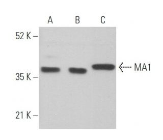
MA1 Antibody (G-10): sc-166915
- MA1 Antibody (G-10) is a mouse monoclonal IgG1 κ MA1 antibody provided at 200 µg/ml
- specific for an epitope mapping between amino acids 67-84 within an internal region of MA1 of mouse origin
- MA1 Antibody (G-10) is recommended for detection of MA1 of mouse, rat and human origin by WB, IP, IF and ELISA
- Anti-MA1 Antibody (G-10) is available conjugated to agarose for IP; HRP for WB, IHC(P) and ELISA; and to either phycoerythrin or FITC for IF, IHC(P) and FCM
- also available conjugated to Alexa Fluor® 488, Alexa Fluor® 546, Alexa Fluor® 594 or Alexa Fluor® 647 for WB (RGB), IF, IHC(P) and FCM, and for use with RGB fluorescent imaging systems, such as iBright™ FL1000, FluorChem™, Typhoon, Azure and other comparable systems
- also available conjugated to Alexa Fluor® 680 or Alexa Fluor® 790 for WB (NIR), IF and FCM; for use with Near-Infrared (NIR) detection systems, such as LI-COR®Odyssey®, iBright™ FL1000, FluorChem™, Typhoon, Azure and other comparable systems
- m-IgG Fc BP-HRP and m-IgGκ BP-HRP are the preferred secondary detection reagents for MA1 Antibody (G-10) for WB applications. These reagents are now offered in bundles with MA1 Antibody (G-10) (see ordering information below).

QUICK LINKS
MA1 Antibody (G-10) is a mouse monoclonal IgG1 kappa light chain antibody that detects MA1 of mouse, rat, and human origin by western blotting (WB), immunoprecipitation (IP), immunofluorescence (IF), and enzyme-linked immunosorbent assay (ELISA). MA1 (G-10) antibody is available in both non-conjugated and various conjugated forms, including agarose, horseradish peroxidase (HRP), phycoerythrin (PE), fluorescein isothiocyanate (FITC), and multiple Alexa Fluor® conjugates. MA1, also known as neuron- and testis-specific protein 1, plays a crucial role in the cellular response to stress and is primarily localized in the nucleolus of normal cells. However, in tumor cells, MA1 translocates to the cytoplasm, which is significant as this relocation is often associated with paraneoplastic neurological disorders (PNDs). PNDs are rare syndromes linked to underlying neoplasms, with testicular cancer being the most common among young males and lung cancer prevalent in other demographics. The immune response against onconeural antigens, such as MA1, can lead to progressive neurological damage, highlighting the importance of understanding MA1 function and localization in both normal and cancerous cells. The MA family includes three known members: MA1, MA2, and MA3, with MA2 being expressed in the brain and testis, and MA3 showing expression in the brain, testis, heart, trachea, and kidney. MA1 (G-10) antibody′s availability in various forms makes this reagent a versatile tool for researchers studying the implications of MA1 in cancer and neurological disorders.
Alexa Fluor® is a trademark of Molecular Probes Inc., OR., USA
LI-COR® and Odyssey® are registered trademarks of LI-COR Biosciences
MA1 Antibody (G-10) References:
- Ma1, a novel neuron- and testis-specific protein, is recognized by the serum of patients with paraneoplastic neurological disorders. | Dalmau, J., et al. 1999. Brain. 122 (Pt 1): 27-39. PMID: 10050892
- Molecular and clinical diversity in paraneoplastic immunity to Ma proteins. | Rosenfeld, MR., et al. 2001. Ann Neurol. 50: 339-48. PMID: 11558790
- Modelling paraneoplastic CNS disease: T-cells specific for the onconeuronal antigen PNMA1 mediate autoimmune encephalomyelitis in the rat. | Pellkofer, H., et al. 2004. Brain. 127: 1822-30. PMID: 15201193
- The human PNMA family: novel neuronal proteins implicated in paraneoplastic neurological disease. | Schüller, M., et al. 2005. J Neuroimmunol. 169: 172-6. PMID: 16214224
Ordering Information
| Product Name | Catalog # | UNIT | Price | Qty | FAVORITES | |
MA1 Antibody (G-10) | sc-166915 | 200 µg/ml | $322.00 | |||
MA1 Antibody (G-10): m-IgG Fc BP-HRP Bundle | sc-529085 | 200 µg Ab; 10 µg BP | $361.00 | |||
MA1 Antibody (G-10): m-IgGκ BP-HRP Bundle | sc-521797 | 200 µg Ab, 40 µg BP | $361.00 | |||
MA1 Antibody (G-10) AC | sc-166915 AC | 500 µg/ml, 25% agarose | $424.00 | |||
MA1 Antibody (G-10) HRP | sc-166915 HRP | 200 µg/ml | $322.00 | |||
MA1 Antibody (G-10) FITC | sc-166915 FITC | 200 µg/ml | $336.00 | |||
MA1 Antibody (G-10) PE | sc-166915 PE | 200 µg/ml | $349.00 | |||
MA1 Antibody (G-10) Alexa Fluor® 488 | sc-166915 AF488 | 200 µg/ml | $364.00 | |||
MA1 Antibody (G-10) Alexa Fluor® 546 | sc-166915 AF546 | 200 µg/ml | $364.00 | |||
MA1 Antibody (G-10) Alexa Fluor® 594 | sc-166915 AF594 | 200 µg/ml | $364.00 | |||
MA1 Antibody (G-10) Alexa Fluor® 647 | sc-166915 AF647 | 200 µg/ml | $364.00 | |||
MA1 Antibody (G-10) Alexa Fluor® 680 | sc-166915 AF680 | 200 µg/ml | $364.00 | |||
MA1 Antibody (G-10) Alexa Fluor® 790 | sc-166915 AF790 | 200 µg/ml | $364.00 | |||
MA1 (G-10) Neutralizing Peptide | sc-166915 P | 100 µg/0.5 ml | $69.00 |