")
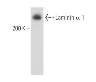
: sc-74418. Western blot analysis of Laminin α-1 expression in Caki-1 whole cell lysate.")
: sc-74418. Western blot analysis of Laminin α-1 expression in Caki-1 whole cell lysate (A) and human testis tissue extract (B). Detection reagent used: m-IgG<sub>1</sub> BP-HRP: sc-525408.")
Laminin α-1 Antibody (G-12): sc-74418
- Laminin α-1 Antibody (G-12) is a mouse monoclonal IgG1 κ Laminin α-1 antibody, cited in 9 publications, provided at 200 µg/ml
- raised against amino acids 1856-2099 mapping within an internal region of Laminin α-1 of human origin
- Laminin alpha-1 Antibody (G-12) is recommended for detection of Laminin α-1 of human origin by WB, IP, IF and ELISA
- Anti-Laminin alpha-1 Antibody (G-12) is available conjugated to agarose for IP; HRP for WB, IHC(P) and ELISA; and to either phycoerythrin or FITC for IF, IHC(P) and FCM
- also available conjugated to Alexa Fluor® 488, Alexa Fluor® 546, Alexa Fluor® 594 or Alexa Fluor® 647 for WB (RGB), IF, IHC(P) and FCM, and for use with RGB fluorescent imaging systems, such as iBright™ FL1000, FluorChem™, Typhoon, Azure and other comparable systems
- also available conjugated to Alexa Fluor® 680 or Alexa Fluor® 790 for WB (NIR), IF and FCM; for use with Near-Infrared (NIR) detection systems, such as LI-COR®Odyssey®, iBright™ FL1000, FluorChem™, Typhoon, Azure and other comparable systems
- m-IgG Fc BP-HRP and m-IgG1 BP-HRP are the preferred secondary detection reagents for Laminin α-1 Antibody (G-12) for WB applications. These reagents are now offered in bundles with Laminin α-1 Antibody (G-12) (see ordering information below).

QUICK LINKS
SEE ALSO...
Laminin α-1 Antibody (G-12) is a mouse monoclonal IgG1 kappa light chain antibody that detects Laminin α-1 protein of human origin by western blotting (WB), immunoprecipitation (IP), immunofluorescence (IF), and enzyme-linked immunosorbent assay (ELISA). Laminin α-1 (G-12) antibody is available in both non-conjugated and various conjugated forms, including agarose, horseradish peroxidase (HRP), phycoerythrin (PE), fluorescein isothiocyanate (FITC), and multiple Alexa Fluor® conjugates. Laminin α-1 is a crucial component of basement membranes, which are specialized extracellular matrices that provide structural support to tissues and play a vital role in cell signaling and tissue organization. These membranes are primarily located beneath epithelial cells, surrounding blood vessels, and enveloping muscle and nerve cells, thereby influencing processes such as cell adhesion, migration, and differentiation. The unique structure of laminins, which consists of three polypeptide chains (α, β, and γ), allows them to form a network that interacts with other extracellular matrix components, facilitating the assembly of basement membranes. Laminin α-1 has been implicated in various pathological conditions, including overexpression in the frontal cortex of patients with Alzheimer′s disease, highlighting a potential role in neurodegenerative processes. The human Laminin α-1 gene is located on chromosome 18p11.3, and understanding its function and interactions is essential for elucidating contributions to both normal physiology and disease states.
Alexa Fluor® is a trademark of Molecular Probes Inc., OR., USA
LI-COR® and Odyssey® are registered trademarks of LI-COR Biosciences
Laminin α-1 Antibody (G-12) References:
- Role of laminin isoforms in glomerular structure. | Hansen, K. and Abrass, CK. 1999. Pathobiology. 67: 84-91. PMID: 10023136
- Laminin alpha chains in colon carcinoma cell lines: detection of a truncated laminin alpha1 mRNA in Caco-2 cells. | Velling, T., et al. 1999. Exp Cell Res. 248: 627-33. PMID: 10222155
- Form and function: the laminin family of heterotrimers. | Colognato, H. and Yurchenco, PD. 2000. Dev Dyn. 218: 213-34. PMID: 10842354
- Transcriptional regulation of laminin gene expression. | Aberdam, D., et al. 2000. Microsc Res Tech. 51: 228-37. PMID: 11054873
- Identification of homologous biologically active sites on the N-terminal domain of laminin alpha chains. | Nomizu, M., et al. 2001. Biochemistry. 40: 15310-7. PMID: 11735413
- Biological activities of the homologous loop regions in the laminin α chain LG modules. | Katagiri, F., et al. 2014. Biochemistry. 53: 3699-708. PMID: 24850085
- Differential proteolytic susceptibility of laminin alpha and beta subunits. | Rao, CN., et al. 1982. Arch Biochem Biophys. 219: 65-70. PMID: 6758704
- The laminin family. | Tryggvason, K. 1993. Curr Opin Cell Biol. 5: 877-82. PMID: 8240830
- Role of laminin in endothelial cell recognition and differentiation. | Schnaper, HW., et al. 1993. Kidney Int. 43: 20-5. PMID: 8433560
- Domains of laminin. | Engvall, E. and Wewer, UM. 1996. J Cell Biochem. 61: 493-501. PMID: 8806072
- Laminin and the mechanism of neuronal outgrowth. | Luckenbill-Edds, L. 1997. Brain Res Brain Res Rev. 23: 1-27. PMID: 9063584
- Laminin isoforms and epithelial development. | Ekblom, M., et al. 1998. Ann N Y Acad Sci. 857: 194-211. PMID: 9917842
Ordering Information
| Product Name | Catalog # | UNIT | Price | Qty | FAVORITES | |
Laminin α-1 Antibody (G-12) | sc-74418 | 200 µg/ml | $322.00 | |||
Laminin α-1 Antibody (G-12): m-IgG Fc BP-HRP Bundle | sc-527018 | 200 µg Ab; 10 µg BP | $361.00 | |||
Laminin α-1 Antibody (G-12): m-IgG1 BP-HRP Bundle | sc-532391 | 200 µg Ab; 20 µg BP | $361.00 | |||
Laminin α-1 Antibody (G-12) AC | sc-74418 AC | 500 µg/ml, 25% agarose | $424.00 | |||
Laminin α-1 Antibody (G-12) HRP | sc-74418 HRP | 200 µg/ml | $322.00 | |||
Laminin α-1 Antibody (G-12) FITC | sc-74418 FITC | 200 µg/ml | $336.00 | |||
Laminin α-1 Antibody (G-12) PE | sc-74418 PE | 200 µg/ml | $349.00 | |||
Laminin α-1 Antibody (G-12) Alexa Fluor® 488 | sc-74418 AF488 | 200 µg/ml | $364.00 | |||
Laminin α-1 Antibody (G-12) Alexa Fluor® 546 | sc-74418 AF546 | 200 µg/ml | $364.00 | |||
Laminin α-1 Antibody (G-12) Alexa Fluor® 594 | sc-74418 AF594 | 200 µg/ml | $364.00 | |||
Laminin α-1 Antibody (G-12) Alexa Fluor® 647 | sc-74418 AF647 | 200 µg/ml | $364.00 | |||
Laminin α-1 Antibody (G-12) Alexa Fluor® 680 | sc-74418 AF680 | 200 µg/ml | $364.00 | |||
Laminin α-1 Antibody (G-12) Alexa Fluor® 790 | sc-74418 AF790 | 200 µg/ml | $364.00 |