")
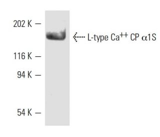
: sc-21782. Western blot analysis of L-type Ca<sup>++</sup> CP α1S expression in rabbit KCl-treated microsomes. Kindly provided by L. McDonough at University of Iowa.")
L-type Ca++ CP α1S Antibody (IIF71VH3): sc-21782
- L-type Ca++ CP α1S Antibody (IIF71VH3) is a mouse monoclonal IgG1, cited in 2 publications, provided at 200 µg/ml
- raised against rabbit skeletal muscle triads
- recommended for detection of 170 kDa, L-type calcium channel α1S of mouse, rat, human and rabbit origin by WB, IP and IF
- At present, we have not yet completed the identification of the preferred secondary detection reagent(s) for L-type Ca++ CP α1S Antibody (IIF71VH3). This work is in progress.

QUICK LINKS
SEE ALSO...
L-type Ca++ CP α1S Antibody (IIF71VH3) is a mouse monoclonal IgG1 antibody that detects L-type Ca++ CP α1S in mouse, rat, human, and rabbit samples through applications such as western blotting (WB), immunoprecipitation (IP), and immunofluorescence (IF). L-type Ca++ CP α1S is a critical component of voltage-dependent calcium channels, which are essential for mediating calcium entry into excitable cells in response to membrane depolarization. These channels play a pivotal role in various calcium-dependent processes, including muscle contraction, hormone and neurotransmitter release, and gene expression. The structure of L-type calcium channels consists of multimeric complexes composed of an α-1 subunit, an intracellular β-subunit, a disulfide-linked α-2/δ subunit, and a transmembrane γ-subunit. The α-1S subunit specifically is crucial for excitation-contraction coupling in skeletal muscle, facilitating the conversion of electrical signals into mechanical action. Additionally, L-type calcium channels can form macromolecular signaling complexes with G protein-coupled receptors, enhancing the specificity of signaling pathways that regulate muscle function and other cellular processes. The diverse biophysical and pharmacologic properties of these channels, including their classification into various types such as L, N, T, P, Q, and R, underscore their importance in cellular physiology and highlight the value of anti-L-type Ca++ CP α1S antibody (IIF71VH3) in research focused on calcium signaling and muscle physiology.
Alexa Fluor® is a trademark of Molecular Probes Inc., OR., USA
LI-COR® and Odyssey® are registered trademarks of LI-COR Biosciences
L-type Ca++ CP α1S Antibody (IIF71VH3) References:
- Regulation of muscle Cav1.1 channels by long-term depolarization involves proteolysis of the alpha1s subunit. | Carrillo, E., et al. 2004. J Membr Biol. 199: 155-61. PMID: 15457372
- The junctional SR protein JP-45 affects the functional expression of the voltage-dependent Ca2+ channel Cav1.1. | Anderson, AA., et al. 2006. J Cell Sci. 119: 2145-55. PMID: 16638807
- Sequence differences in the IQ motifs of CaV1.1 and CaV1.2 strongly impact calmodulin binding and calcium-dependent inactivation. | Ohrtman, J., et al. 2008. J Biol Chem. 283: 29301-11. PMID: 18718913
- A CaV1.1 Ca2+ channel splice variant with high conductance and voltage-sensitivity alters EC coupling in developing skeletal muscle. | Tuluc, P., et al. 2009. Biophys J. 96: 35-44. PMID: 19134469
- Beneficial effects of bumetanide in a CaV1.1-R528H mouse model of hypokalaemic periodic paralysis. | Wu, F., et al. 2013. Brain. 136: 3766-74. PMID: 24142145
- Ca(2+) permeation and/or binding to CaV1.1 fine-tunes skeletal muscle Ca(2+) signaling to sustain muscle function. | Lee, CS., et al. 2015. Skelet Muscle. 5: 4. PMID: 25717360
- Ca2+ Binding/Permeation via Calcium Channel, CaV1.1, Regulates the Intracellular Distribution of the Fatty Acid Transport Protein, CD36, and Fatty Acid Metabolism. | Georgiou, DK., et al. 2015. J Biol Chem. 290: 23751-65. PMID: 26245899
- T Cell Receptor Mediated Calcium Entry Requires Alternatively Spliced Cav1.1 Channels. | Matza, D., et al. 2016. PLoS One. 11: e0147379. PMID: 26815481
- Calpain inhibition rescues troponin T3 fragmentation, increases Cav1.1, and enhances skeletal muscle force in aging sedentary mice. | Zhang, T., et al. 2016. Aging Cell. 15: 488-98. PMID: 26892246
- Two distinct voltage-sensing domains control voltage sensitivity and kinetics of current activation in CaV1.1 calcium channels. | Tuluc, P., et al. 2016. J Gen Physiol. 147: 437-49. PMID: 27185857
Ordering Information
| Product Name | Catalog # | UNIT | Price | Qty | FAVORITES | |
L-type Ca++ CP α1S Antibody (IIF71VH3) | sc-21782 | 200 µg/ml | $322.00 |