")
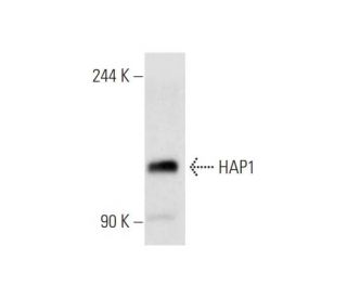
: sc-166245. Western blot analysis of HAP1 expression in COLO 320DM whole cell lysate.")
: sc-166245. Immunoperoxidase staining of formalin fixed, paraffin-embedded human cerebral cortex tissue showing cytoplasmic staining of neuronal cells and glial cells.")
: sc-166245. Western blot analysis of HAP1 expression in Neuro-2A whole cell lysate.")
HAP1 Antibody (C-3): sc-166245
- HAP1 Antibody (C-3) is a mouse monoclonal IgG1 κ HAP1 antibody, cited in 1 publications, provided at 200 µg/ml
- raised against amino acids 329-628 mapping at the C-terminus of HAP1B of mouse origin
- HAP1 Antibody (C-3) is recommended for detection of HAP1 of mouse, rat and human origin by WB, IP, IF, IHC(P) and ELISA
- Anti-HAP1 Antibody (C-3) is available conjugated to agarose for IP; HRP for WB, IHC(P) and ELISA; and to either phycoerythrin or FITC for IF, IHC(P) and FCM
- also available conjugated to Alexa Fluor® 488, Alexa Fluor® 546, Alexa Fluor® 594 or Alexa Fluor® 647 for WB (RGB), IF, IHC(P) and FCM, and for use with RGB fluorescent imaging systems, such as iBright™ FL1000, FluorChem™, Typhoon, Azure and other comparable systems
- also available conjugated to Alexa Fluor® 680 or Alexa Fluor® 790 for WB (NIR), IF and FCM; for use with Near-Infrared (NIR) detection systems, such as LI-COR®Odyssey®, iBright™ FL1000, FluorChem™, Typhoon, Azure and other comparable systems
- m-IgG Fc BP-HRP and m-IgG1 BP-HRP are the preferred secondary detection reagents for HAP1 Antibody (C-3) for WB and IHC(P) applications. These reagents are now offered in bundles with HAP1 Antibody (C-3) (see ordering information below).

QUICK LINKS
HAP1 Antibody (C-3) is a mouse monoclonal IgG1 kappa light chain antibody that detects HAP1 protein of mouse, rat, and human origin by western blotting (WB), immunoprecipitation (IP), immunofluorescence (IF), immunohistochemistry, and enzyme-linked immunosorbent assay (ELISA). HAP1 Antibody (C-3) is available in non-conjugated and various conjugated forms, including agarose, horseradish peroxidase (HRP), phycoerythrin (PE), fluorescein isothiocyanate (FITC), and multiple Alexa Fluor® conjugates. HAP1, or huntingtin-associated protein 1, plays a crucial role in neuronal function by binding to huntingtin, a protein containing a polyglutamine region. An expanded polyglutamine repeat in huntingtin exceeding 35 repeats is associated with Huntington′s disease, a neurodegenerative disorder. HAP1′s binding to huntingtin increases in the presence of this expanded region, highlighting HAP1′s potential role in disease pathogenesis. HAP1 exhibits distinct neuronal localization, moving along nerve fibers with huntingtin, and is primarily expressed in brain tissue, particularly in the olfactory bulb and brain stem. In rat models, HAP1 associates with various intracellular organelles, while in mouse neurons, HAP1 localizes to membrane-bound organelles, including large endosomes, tubulovesicular structures, and budding vesicles, emphasizing HAP1′s importance in intracellular transport and signaling pathways.
Alexa Fluor® is a trademark of Molecular Probes Inc., OR., USA
LI-COR® and Odyssey® are registered trademarks of LI-COR Biosciences
HAP1 Antibody (C-3) References:
- Nuclear and neuropil aggregates in Huntington's disease: relationship to neuropathology. | Gutekunst, CA., et al. 1999. J Neurosci. 19: 2522-34. PMID: 10087066
- A huntingtin-associated protein enriched in brain with implications for pathology. | Li, XJ., et al. 1995. Nature. 378: 398-402. PMID: 7477378
- A novel gene containing a trinucleotide repeat that is expanded and unstable on Huntington's disease chromosomes. The Huntington's Disease Collaborative Research Group. | . 1993. Cell. 72: 971-83. PMID: 8458085
- Huntingtin-associated protein (HAP1): discrete neuronal localizations in the brain resemble those of neuronal nitric oxide synthase. | Li, XJ., et al. 1996. Proc Natl Acad Sci U S A. 93: 4839-44. PMID: 8643490
- Fast transport and retrograde movement of huntingtin and HAP 1 in axons. | Block-Galarza, J., et al. 1997. Neuroreport. 8: 2247-51. PMID: 9243620
- Huntington's disease. | Gusella, JF., et al. 1996. Cold Spring Harb Symp Quant Biol. 61: 615-26. PMID: 9246488
- Analysis of Huntingtin-associated protein 1 in mouse brain and immortalized striatal neurons. | Martin, EJ., et al. 1999. J Comp Neurol. 403: 421-30. PMID: 9888310
Ordering Information
| Product Name | Catalog # | UNIT | Price | Qty | FAVORITES | |
HAP1 Antibody (C-3) | sc-166245 | 200 µg/ml | $322.00 | |||
HAP1 Antibody (C-3): m-IgG Fc BP-HRP Bundle | sc-527147 | 200 µg Ab; 10 µg BP | $361.00 | |||
HAP1 Antibody (C-3): m-IgG1 BP-HRP Bundle | sc-532520 | 200 µg Ab; 20 µg BP | $361.00 | |||
HAP1 Antibody (C-3) AC | sc-166245 AC | 500 µg/ml, 25% agarose | $424.00 | |||
HAP1 Antibody (C-3) HRP | sc-166245 HRP | 200 µg/ml | $322.00 | |||
HAP1 Antibody (C-3) FITC | sc-166245 FITC | 200 µg/ml | $336.00 | |||
HAP1 Antibody (C-3) PE | sc-166245 PE | 200 µg/ml | $349.00 | |||
HAP1 Antibody (C-3) Alexa Fluor® 488 | sc-166245 AF488 | 200 µg/ml | $364.00 | |||
HAP1 Antibody (C-3) Alexa Fluor® 546 | sc-166245 AF546 | 200 µg/ml | $364.00 | |||
HAP1 Antibody (C-3) Alexa Fluor® 594 | sc-166245 AF594 | 200 µg/ml | $364.00 | |||
HAP1 Antibody (C-3) Alexa Fluor® 647 | sc-166245 AF647 | 200 µg/ml | $364.00 | |||
HAP1 Antibody (C-3) Alexa Fluor® 680 | sc-166245 AF680 | 200 µg/ml | $364.00 | |||
HAP1 Antibody (C-3) Alexa Fluor® 790 | sc-166245 AF790 | 200 µg/ml | $364.00 |