")
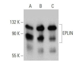
EPLIN Antibody (20): sc-136399
- EPLIN Antibody (20) is a mouse monoclonal IgG1 κ EPLIN antibody, cited in 13 publications, provided at 200 µg/ml
- raised against amino acids 1-141 corresponding to the α isoform of EPLIN of human origin
- recommended for detection of EPLIN of mouse, rat, human and canine origin by WB, IP, IF and IHC(P)
- available conjugated to agarose for IP; and to HRP for WB, IHC(P) and ELISA
- m-IgG Fc BP-HRP, m-IgG1 BP-HRP and m-IgGκ BP-HRP are the preferred secondary detection reagents for EPLIN Antibody (20) for WB and IHC(P) applications. These reagents are now offered in bundles with EPLIN Antibody (20) (see ordering information below).

QUICK LINKS
SEE ALSO...
EPLIN Antibody (20) is a mouse monoclonal IgG1 kappa light chain antibody that detects EPLIN protein of mouse, rat, human, and canine origin by western blotting (WB), immunoprecipitation (IP), immunofluorescence (IF), and immunohistochemistry with paraffin-embedded sections (IHCP). EPLIN Antibody (20) is available in both non-conjugated and various conjugated forms, including agarose and horseradish peroxidase (HRP). Epithelial protein lost in neoplasm (EPLIN) plays a crucial role in regulating the actin cytoskeleton, which is essential for maintaining cell shape, motility, and integrity. EPLIN is characterized by a single centrally located lin-11, isl-1, and mec-3 (LIM) domain, along with at least two actin-binding domains, where the C-terminal domain exhibits stronger binding affinity than the N-terminal domain. By interacting with actin monomers and filaments, EPLIN enhances formation and stability of actin stress fibers, delays filament nucleation, and reduces formation of branched filaments, thereby promoting bundling of actin filaments. EPLIN inhibits membrane ruffling and prevents actin filament depolymerization, which is vital for cellular processes such as migration and adhesion. EPLIN expression is notably high in tissues such as placenta, kidney, pancreas, prostate, ovary, spleen, and heart, while lower levels are found in lung, liver, brain, skeletal muscle, thymus, testis, and intestine. EPLIN exists in two isoforms, EPLIN-alpha and EPLIN-beta, with downregulation of EPLIN-alpha being linked to increased motility of invasive tumor cells, highlighting its potential role in cancer progression.
Alexa Fluor® is a trademark of Molecular Probes Inc., OR., USA
LI-COR® and Odyssey® are registered trademarks of LI-COR Biosciences
EPLIN Antibody (20) References:
- EPLIN, epithelial protein lost in neoplasm. | Maul, RS. and Chang, DD. 1999. Oncogene. 18: 7838-41. PMID: 10618726
- Characterization of the human EPLIN (Epithelial Protein Lost in Neoplasm) gene reveals distinct promoters for the two EPLIN isoforms. | Chen, S., et al. 2000. Gene. 248: 69-76. PMID: 10806352
- Characterization of mouse epithelial protein lost in neoplasm (EPLIN) and comparison of mammalian and zebrafish EPLIN. | Maul, RS., et al. 2001. Gene. 262: 155-60. PMID: 11179679
- Inhibition of anchorage-independent growth of transformed NIH3T3 cells by epithelial protein lost in neoplasm (EPLIN) requires localization of EPLIN to actin cytoskeleton. | Song, Y., et al. 2002. Mol Biol Cell. 13: 1408-16. PMID: 11950948
- EPLIN regulates actin dynamics by cross-linking and stabilizing filaments. | Maul, RS., et al. 2003. J Cell Biol. 160: 399-407. PMID: 12566430
- Compositional characterization of the cytoskeleton of NK-like cells. | Meng, X. and Wilkins, JA. 2005. J Proteome Res. 4: 2081-7. PMID: 16335953
- EPLIN is a crucial regulator for extrusion of RasV12-transformed cells. | Ohoka, A., et al. 2015. J Cell Sci. 128: 781-9. PMID: 25609711
- EPLIN Expression in Gastric Cancer and Impact on Prognosis and Chemoresistance. | Gong, W., et al. 2021. Biomolecules. 11: PMID: 33917939
- Epithelial Protein Lost in Neoplasm, EPLIN, the Cellular and Molecular Prospects in Cancers. | Zeng, J., et al. 2021. Biomolecules. 11: PMID: 34356662
- EPLIN, a Putative Tumour Suppressor in Colorectal Cancer, Implications in Drug Resistance. | Zeng, J., et al. 2022. Int J Mol Sci. 23: PMID: 36499558
- EPLIN-β is a novel substrate of ornithine decarboxylase antizyme 1 and mediates cellular migration. | Li, D., et al. 2023. J Cell Sci. 136: PMID: 37325974
- The concerted action of SEPT9 and EPLIN modulates the adhesion and migration of human fibroblasts. | Hecht, M., et al. 2024. Life Sci Alliance. 7: PMID: 38719752
Ordering Information
| Product Name | Catalog # | UNIT | Price | Qty | FAVORITES | |
EPLIN Antibody (20) | sc-136399 | 200 µg/ml | $322.00 | |||
EPLIN Antibody (20): m-IgG Fc BP-HRP Bundle | sc-528821 | 200 µg Ab; 10 µg BP | $361.00 | |||
EPLIN Antibody (20): m-IgGκ BP-HRP Bundle | sc-521365 | 200 µg Ab, 40 µg BP | $361.00 | |||
EPLIN Antibody (20): m-IgG1 BP-HRP Bundle | sc-543152 | 200 µg Ab; 20 µg BP | $361.00 | |||
EPLIN Antibody (20) AC | sc-136399 AC | 500 µg/ml, 25% agarose | $424.00 | |||
EPLIN Antibody (20) HRP | sc-136399 HRP | 200 µg/ml | $322.00 |