")
EDG-1/S1P1/S1PR1 Antibody (A-6): sc-48356
- EDG-1/S1P1/S1PR1 Antibody (A-6) is a mouse monoclonal IgG3 κ EDG-1/S1P1/S1PR1 antibody, cited in 23 publications, provided at 200 µg/ml
- raised against amino acids 322-381 of EDG-1 of human origin
- recommended for detection of EDG-1 of human origin by WB, IP, IF, IHC(P) and ELISA
- available conjugated to agarose for IP; HRP for WB, IHC(P) and ELISA; phycoerythrin, FITC, Alexa Fluor® 488 or Alexa Fluor® 647 for IF, IHC(P) and FCM
- At present, we have not yet completed the identification of the preferred secondary detection reagent(s) for EDG-1/S1P1/S1PR1 Antibody (A-6). This work is in progress.

QUICK LINKS
SEE ALSO...
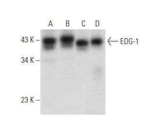
EDG-1 Antibody (A-6) is a mouse monoclonal IgG3 kappa light chain antibody that detects EDG-1 of human origin by western blotting (WB), immunoprecipitation (IP), immunofluorescence (IF), immunohistochemistry, and enzyme-linked immunosorbent assay (ELISA). EDG-1 (A-6) antibody is available in both non-conjugated and various conjugated forms, including agarose, horseradish peroxidase (HRP), phycoerythrin (PE), fluorescein isothiocyanate (FITC), and multiple Alexa Fluor® conjugates. EDG-1, also known as S1PR1 or S1P1, is a member of the endothelial differentiation gene (EDG) family of G protein-coupled receptors, which play a crucial role in mediating the effects of lysophospholipid signaling molecules such as sphingosine-1-phosphate (S1P) and lysophosphatidic acid (LPA). EDG-1 is primarily located on the cell surface and is involved in various cellular processes, including cell survival, growth, migration, and differentiation, by coupling to Gi proteins to activate downstream signaling pathways like Akt. This is particularly important in glioma cells, where EDG-1 expression is associated with tumor progression and survival. EDG-1′s ability to interact with multiple G proteins influences a wide range of physiological responses, making EDG-1 a significant target for research in cancer biology and therapeutic development.
Alexa Fluor® is a trademark of Molecular Probes Inc., OR., USA
LI-COR® and Odyssey® are registered trademarks of LI-COR Biosciences
EDG-1/S1P1/S1PR1 Antibody (A-6) References:
- Differential pharmacological properties and signal transduction of the sphingosine 1-phosphate receptors EDG-1, EDG-3, and EDG-5. | Ancellin, N. and Hla, T. 1999. J Biol Chem. 274: 18997-9002. PMID: 10383399
- A subfamily of G protein-coupled cellular receptors for lysophospholipids and lysosphingolipids. | Goetzl, EJ. and An, S. 1999. Adv Exp Med Biol. 469: 259-64. PMID: 10667339
- Sphingosine-1-phosphate is a ligand for the G protein-coupled receptor EDG-6. | Van Brocklyn, JR., et al. 2000. Blood. 95: 2624-9. PMID: 10753843
- Sphingosine-1-phosphate signaling via the EDG-1 family of G-protein-coupled receptors. | Hla, T., et al. 2000. Ann N Y Acad Sci. 905: 16-24. PMID: 10818438
- Sphingosine 1-phosphate: a ligand for the EDG-1 family of G-protein-coupled receptors. | Spiegel, S. 2000. Ann N Y Acad Sci. 905: 54-60. PMID: 10818441
- Sphingosine 1-phosphate signalling in mammalian cells. | Pyne, S. and Pyne, NJ. 2000. Biochem J. 349: 385-402. PMID: 10880336
- Differential roles of Edg-1 and Edg-5, sphingosine 1-phosphate receptors, in the signaling pathways in C6 glioma cells. | Sato, K., et al. 2000. Brain Res Mol Brain Res. 85: 151-60. PMID: 11146117
- Lysophosphatidic acid receptor-selective effects on Jurkat T cell migration through a Matrigel model basement membrane. | Zheng, Y., et al. 2001. J Immunol. 166: 2317-22. PMID: 11160288
- Edg2 receptor distribution in adult rat brain. | Handford, EJ., et al. 2001. Neuroreport. 12: 757-60. PMID: 11277579
- Sphingosine 1-phosphate activates Akt, nitric oxide production, and chemotaxis through a Gi protein/phosphoinositide 3-kinase pathway in endothelial cells. | Morales-Ruiz, M., et al. 2001. J Biol Chem. 276: 19672-7. PMID: 11278592
- Roles for N-glycosylation in the dynamics of Edg-1/S1P1 in sphingosine 1-phosphate-stimulated cells. | Kohno, T. and Igarashi, Y. 2004. Glycoconj J. 21: 497-501. PMID: 15750791
Ordering Information
| Product Name | Catalog # | UNIT | Price | Qty | FAVORITES | |
EDG-1/S1P1/S1PR1 Antibody (A-6) | sc-48356 | 200 µg/ml | $322.00 | |||
EDG-1/S1P1/S1PR1 Antibody (A-6) AC | sc-48356 AC | 500 µg/ml, 25% agarose | $424.00 | |||
EDG-1/S1P1/S1PR1 Antibody (A-6) Alexa Fluor® 488 | sc-48356 AF488 | 200 µg/ml | $364.00 | |||
EDG-1/S1P1/S1PR1 Antibody (A-6) Alexa Fluor® 647 | sc-48356 AF647 | 200 µg/ml | $364.00 | |||
EDG-1/S1P1/S1PR1 Antibody (A-6) FITC | sc-48356 FITC | 200 µg/ml | $336.00 | |||
EDG-1/S1P1/S1PR1 Antibody (A-6) HRP | sc-48356 HRP | 200 µg/ml | $322.00 | |||
EDG-1/S1P1/S1PR1 Antibody (A-6) PE | sc-48356 PE | 200 µg/ml | $349.00 |