")
Cytokeratin 6/75 Antibody (H-6): sc-166074
- Cytokeratin 6/75 Antibody (H-6) is a mouse monoclonal IgG2a κ Cytokeratin 6/75 antibody, cited in 2 publications, provided at 200 µg/ml
- specific for an epitope mapping between amino acids 147-180 within an internal region of Cytokeratin 6A of human origin
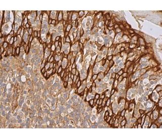
- Cytokeratin 6/75 Antibody (H-6) is recommended for detection of Cytokeratin 6A, Cytokeratin 6B, Cytokeratin 6C and Cytokeratin 75 of mouse, rat and human origin by WB, IP, IF, IHC(P) and ELISA
- Anti-Cytokeratin 6/75 Antibody (H-6) is available conjugated to agarose for IP; HRP for WB, IHC(P) and ELISA; and to either phycoerythrin or FITC for IF, IHC(P) and FCM
- also available conjugated to Alexa Fluor® 488, Alexa Fluor® 546, Alexa Fluor® 594 or Alexa Fluor® 647 for WB (RGB), IF, IHC(P) and FCM, and for use with RGB fluorescent imaging systems, such as iBright™ FL1000, FluorChem™, Typhoon, Azure and other comparable systems
- also available conjugated to Alexa Fluor® 680 or Alexa Fluor® 790 for WB (NIR), IF and FCM; for use with Near-Infrared (NIR) detection systems, such as LI-COR®Odyssey®, iBright™ FL1000, FluorChem™, Typhoon, Azure and other comparable systems
- m-IgG Fc BP-HRP, m-IgG2a BP-HRP and m-IgGκ BP-HRP are the preferred secondary detection reagents for Cytokeratin 6/75 Antibody (H-6) for WB and IHC(P) applications. These reagents are now offered in bundles with Cytokeratin 6/75 Antibody (H-6) (see ordering information below).

QUICK LINKS
SEE ALSO...
Cytokeratin 6/75 Antibody (H-6) is a mouse monoclonal IgG2a kappa light chain antibody that detects Cytokeratin 6/75 protein of mouse, rat, and human origin by western blotting (WB), immunoprecipitation (IP), immunofluorescence (IF), immunohistochemistry with paraffin-embedded sections (IHCP), and enzyme-linked immunosorbent assay (ELISA). Anti-Cytokeratin 6/75 antibody (H-6) is available in both non-conjugated and various conjugated forms, including agarose, horseradish peroxidase (HRP), phycoerythrin (PE), fluorescein isothiocyanate (FITC), and multiple Alexa Fluor® conjugates. Cytokeratin 6/75 is a member of the cytokeratin family, consisting of diverse intermediate filament proteins crucial for maintaining epithelial cell structural integrity. These proteins are expressed in pairs within keratinized and non-keratinized epithelial tissues, constituting up to 85% of mature keratinocytes in vertebrate epidermis. α-helical coiled-coil dimers associate laterally to form 10 nm diameter filaments, providing mechanical support and resilience to epithelial tissues. Cytokeratin 6A is predominantly found in epithelial tissues, highlighting importance in tissue differentiation and specialization. Human Cytokeratin 6A gene is located on chromosome 12q13, and mutations are associated with several inheritable hair and skin disorders. Cytokeratin 75, also known as K6HF, maps to the same genetic locus and plays a significant role in the companion layer of hair follicles, emphasizing importance in both normal physiology and disease states.
Alexa Fluor® is a trademark of Molecular Probes Inc., OR., USA
LI-COR® and Odyssey® are registered trademarks of LI-COR Biosciences
Cytokeratin 6/75 Antibody (H-6) References:
- Identification of sporadic mutations in the helix initiation motif of keratin 6 in two pachyonychia congenita patients: further evidence for a mutational hot spot. | Lin, MT., et al. 1999. Exp Dermatol. 8: 115-9. PMID: 10232401
- Monilethrix: mutational hotspot in the helix termination motif of the human hair basic keratin 6. | Horev, L., et al. 2000. Hum Hered. 50: 325-30. PMID: 10878479
- Inflammatory versus proliferative processes in epidermis. Tumor necrosis factor alpha induces K6b keratin synthesis through a transcriptional complex containing NFkappa B and C/EBPbeta. | Komine, M., et al. 2000. J Biol Chem. 275: 32077-88. PMID: 10887174
- Interleukin-1 induces transcription of keratin K6 in human epidermal keratinocytes. | Komine, M., et al. 2001. J Invest Dermatol. 116: 330-8. PMID: 11180011
- Three epidermal and one simple epithelial type II keratin genes map to human chromosome 12. | Rosenberg, M., et al. 1991. Cytogenet Cell Genet. 57: 33-8. PMID: 1713141
- Cytokeratin 5 and cytokeratin 6 expressions are unconnected in normal and cancerous tissues and have separate diagnostic implications. | Völkel, C., et al. 2022. Virchows Arch. 480: 433-447. PMID: 34559291
- Cytokeratin 6 identifies basal-like subtypes of pancreatic ductal adenocarcinoma with decreased survival. | Lyu, SI., et al. 2023. J Cancer Res Clin Oncol. 149: 7539-7546. PMID: 36971797
- Cloning and characterization of multiple human genes and cDNAs encoding highly related type II keratin 6 isoforms. | Takahashi, K., et al. 1995. J Biol Chem. 270: 18581-92. PMID: 7543104
- Keratins and the skin. | Fuchs, E. 1995. Annu Rev Cell Dev Biol. 11: 123-53. PMID: 8689554
- Functional inactivation of p53 by antisense RNA induces invasive ability of lung carcinoma cells and downregulates cytokeratin synthesis. | Mukhopadhyay, T. and Roth, JA. 1996. Anticancer Res. 16: 1683-9. PMID: 8712687
- A mutational hotspot in the 2B domain of human hair basic keratin 6 (hHb6) in monilethrix patients. | Korge, BP., et al. 1998. J Invest Dermatol. 111: 896-9. PMID: 9804356
- A novel human type II cytokeratin, K6hf, specifically expressed in the companion layer of the hair follicle. | Winter, H., et al. 1998. J Invest Dermatol. 111: 955-62. PMID: 9856802
Ordering Information
| Product Name | Catalog # | UNIT | Price | Qty | FAVORITES | |
Cytokeratin 6/75 Antibody (H-6) | sc-166074 | 200 µg/ml | $322.00 | |||
Cytokeratin 6/75 Antibody (H-6): m-IgG Fc BP-HRP Bundle | sc-528902 | 200 µg Ab; 10 µg BP | $361.00 | |||
Cytokeratin 6/75 Antibody (H-6): m-IgGκ BP-HRP Bundle | sc-521503 | 200 µg Ab, 40 µg BP | $361.00 | |||
Cytokeratin 6/75 Antibody (H-6): m-IgG2a BP-HRP Bundle | sc-547203 | 200 µg Ab; 10 µg BP | $361.00 | |||
Cytokeratin 6/75 Antibody (H-6) AC | sc-166074 AC | 500 µg/ml, 25% agarose | $424.00 | |||
Cytokeratin 6/75 Antibody (H-6) HRP | sc-166074 HRP | 200 µg/ml | $322.00 | |||
Cytokeratin 6/75 Antibody (H-6) FITC | sc-166074 FITC | 200 µg/ml | $336.00 | |||
Cytokeratin 6/75 Antibody (H-6) PE | sc-166074 PE | 200 µg/ml | $349.00 | |||
Cytokeratin 6/75 Antibody (H-6) Alexa Fluor® 488 | sc-166074 AF488 | 200 µg/ml | $364.00 | |||
Cytokeratin 6/75 Antibody (H-6) Alexa Fluor® 546 | sc-166074 AF546 | 200 µg/ml | $364.00 | |||
Cytokeratin 6/75 Antibody (H-6) Alexa Fluor® 594 | sc-166074 AF594 | 200 µg/ml | $364.00 | |||
Cytokeratin 6/75 Antibody (H-6) Alexa Fluor® 647 | sc-166074 AF647 | 200 µg/ml | $364.00 | |||
Cytokeratin 6/75 Antibody (H-6) Alexa Fluor® 680 | sc-166074 AF680 | 200 µg/ml | $364.00 | |||
Cytokeratin 6/75 Antibody (H-6) Alexa Fluor® 790 | sc-166074 AF790 | 200 µg/ml | $364.00 | |||
Cytokeratin 6/75 (H-6) Neutralizing Peptide | sc-166074 P | 100 µg/0.5 ml | $69.00 |