")
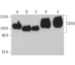
CD43 Antibody (DF-T1): sc-6256
- CD43 Antibody (DF-T1) is a mouse monoclonal IgG1 κ CD43 antibody, cited in 8 publications, provided at 200 µg/ml
- raised against myeloblastic cell line KG1
- CD43 Antibody (DF-T1) is recommended for detection of CD43 of human origin by WB, IP, IF, IHC(P) and FCM
- Anti-CD43 Antibody (DF-T1) is available conjugated to agarose for IP; HRP for WB, IHC(P) and ELISA; and to either phycoerythrin or FITC for IF, IHC(P) and FCM
- also available conjugated to Alexa Fluor® 488, Alexa Fluor® 546, Alexa Fluor® 594 or Alexa Fluor® 647 for WB (RGB), IF, IHC(P) and FCM, and for use with RGB fluorescent imaging systems, such as iBright™ FL1000, FluorChem™, Typhoon, Azure and other comparable systems
- also available conjugated to Alexa Fluor® 680 or Alexa Fluor® 790 for WB (NIR), IF and FCM; for use with Near-Infrared (NIR) detection systems, such as LI-COR®Odyssey®, iBright™ FL1000, FluorChem™, Typhoon, Azure and other comparable systems
- m-IgG Fc BP-HRP, m-IgG1 BP-HRP and m-IgGκ BP-HRP are the preferred secondary detection reagents for CD43 Antibody (DF-T1) for WB and IHC(P) applications. These reagents are now offered in bundles with CD43 Antibody (DF-T1) (see ordering information below).

QUICK LINKS
SEE ALSO...
CD43 Antibody (DF-T1) is a mouse monoclonal IgG1 kappa light chain antibody that detects CD43 protein of human origin by western blotting (WB), immunoprecipitation (IP), immunofluorescence (IF), immunohistochemistry with paraffin-embedded sections (IHCP), and flow cytometry (FCM). CD43 Antibody (DF-T1) is available in both non-conjugated and various conjugated forms, including agarose, horseradish peroxidase (HRP), phycoerythrin (PE), fluorescein isothiocyanate (FITC), and multiple Alexa Fluor® conjugates. CD43, also known as leukosialin, is a major O-glycosylated sialoglycoprotein predominantly found on the surface of leukocytes, playing a crucial role in cellular adhesion and tumor progression. CD43 is a member of the surface mucin family and is characterized by extensive glycosylation, which is essential for function in modulating cell-cell interactions and immune responses. CD43 glycosylation not only influences structural properties but also affects interactions with other proteins, thereby impacting processes such as leukocyte migration and activation. CD43 is particularly important in the context of T cells and certain B cell populations, serving as a key marker for identifying both normal and neoplastic cells in various tissues. By functioning as a negative regulator of cellular adhesion, CD43 helps to maintain the balance of immune responses, making CD43 a significant target for research in immunology and cancer biology.
Alexa Fluor® is a trademark of Molecular Probes Inc., OR., USA
LI-COR® and Odyssey® are registered trademarks of LI-COR Biosciences
CD43 Antibody (DF-T1) References:
- [Phenotypical and functional characterization of leukocytes--the CD-system]. | Holter, W., et al. 1991. Wien Klin Wochenschr. 103: 247-62. PMID: 2068816
- CD43 in the nucleus and cytoplasm of lung cancer is a potential therapeutic target. | Fu, Q., et al. 2013. Int J Cancer. 132: 1761-70. PMID: 23015282
- Cancer-associated CD43 glycoforms as target of immunotherapy. | Tuccillo, FM., et al. 2014. Mol Cancer Ther. 13: 752-62. PMID: 24356816
- Aberrant glycosylation as biomarker for cancer: focus on CD43. | Tuccillo, FM., et al. 2014. Biomed Res Int. 2014: 742831. PMID: 24689054
- CD43 in the malignant flow cytometry laboratory in 2020. | Sorigue, M. 2021. Expert Rev Hematol. 14: 123-136. PMID: 33249940
- CD11/CD18 panel report for swine CD workshop. | Kim, YB., et al. 1994. Vet Immunol Immunopathol. 43: 289-91. PMID: 7531911
- Regulatory role of CD43 leukosialin on integrin-mediated T-cell adhesion to endothelial and extracellular matrix ligands and its polar redistribution to a cellular uropod. | Sánchez-Mateos, P., et al. 1995. Blood. 86: 2228-39. PMID: 7545023
- Negative regulation of T-cell adhesion and activation by CD43. | Manjunath, N., et al. 1995. Nature. 377: 535-8. PMID: 7566153
- Expression of the leukocyte-associated sialoglycoprotein CD43 by a colon carcinoma cell line. | Baeckström, D., et al. 1995. J Biol Chem. 270: 13688-92. PMID: 7775421
- CD43 and CD5 antibodies define four normal and neoplastic B-cell subsets: a three-color flow cytometric study. | Lynch, EF., et al. 1995. Cytometry. 22: 223-31. PMID: 8556954
- Specific monoclonal antibodies against leukocyte-restricted cell surface molecule CD43 react with nonhematopoietic tumor cells. | Santamaría, M., et al. 1996. Cancer Res. 56: 3526-9. PMID: 8758921
- The CD43 130-kD peripheral T-cell activation antigen is downregulated in thymic positive selection. | Ellies, LG., et al. 1996. Blood. 88: 1725-32. PMID: 8781428
Ordering Information
| Product Name | Catalog # | UNIT | Price | Qty | FAVORITES | |
CD43 Antibody (DF-T1) | sc-6256 | 200 µg/ml | $322.00 | |||
CD43 Antibody (DF-T1): m-IgG Fc BP-HRP Bundle | sc-528164 | 200 µg Ab; 10 µg BP | $361.00 | |||
CD43 Antibody (DF-T1): m-IgGκ BP-HRP Bundle | sc-520472 | 200 µg Ab, 40 µg BP | $361.00 | |||
CD43 Antibody (DF-T1): m-IgG1 BP-HRP Bundle | sc-542794 | 200 µg Ab; 20 µg BP | $361.00 | |||
CD43 Antibody (DF-T1) AC | sc-6256 AC | 500 µg/ml, 25% agarose | $424.00 | |||
CD43 Antibody (DF-T1) HRP | sc-6256 HRP | 200 µg/ml | $322.00 | |||
CD43 Antibody (DF-T1) FITC | sc-6256 FITC | 200 µg/ml | $336.00 | |||
CD43 Antibody (DF-T1) PE | sc-6256 PE | 200 µg/ml | $349.00 | |||
CD43 Antibody (DF-T1) Alexa Fluor® 488 | sc-6256 AF488 | 200 µg/ml | $364.00 | |||
CD43 Antibody (DF-T1) Alexa Fluor® 546 | sc-6256 AF546 | 200 µg/ml | $364.00 | |||
CD43 Antibody (DF-T1) Alexa Fluor® 594 | sc-6256 AF594 | 200 µg/ml | $364.00 | |||
CD43 Antibody (DF-T1) Alexa Fluor® 647 | sc-6256 AF647 | 200 µg/ml | $364.00 | |||
CD43 Antibody (DF-T1) Alexa Fluor® 680 | sc-6256 AF680 | 200 µg/ml | $364.00 | |||
CD43 Antibody (DF-T1) Alexa Fluor® 790 | sc-6256 AF790 | 200 µg/ml | $364.00 |