Date published: 2026-3-19

1-800-457-3801

SCBT Portrait Logo
Seach Input

C3 Antibody (A-4): sc-55458

5.0(4)
Write a reviewAsk a question

Datasheets
  • C3 Antibody (A-4) is a mouse monoclonal IgG1 κ, cited in 1 publications, provided at 200 µg/ml
  • raised against amino acids 541-840 of C3 of human origin
  • recommended for detection of C3 of human origin by WB, IP, IF and ELISA
  • See C3 (B-9): sc-28294 for C3 antibody conjugates, including AC, HRP, FITC, PE, Alexa Fluor® 488, 594, 647, 680 and 790.
  • m-IgG Fc BP-HRP, m-IgG2a BP-HRP and m-IgGκ BP-HRP are the preferred secondary detection reagents for C3 Antibody (A-4) for WB applications. These reagents are now offered in bundles with C3 Antibody (A-4) (see ordering information below).
Crispr Promo Banner

QUICK LINKS

SEE ALSO...

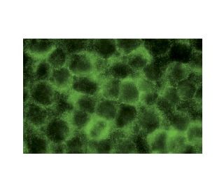
C3 Antibody (A-4) is a mouse monoclonal IgG1 kappa light chain antibody raised against amino acids 541-840 of the human Complement Component 3 (C3) protein. C3, also known as CPAMD1 or acylation-stimulating protein (ASP), is a central component of the complement system, which plays a crucial role in innate immunity. Structurally, C3 is a large glycoprotein composed of an alpha and a beta chain linked by disulfide bonds, forming a complex that is essential for its function in immune defense. A key feature of C3′s structure is the presence of a thioester bond within the alpha chain, which upon activation allows C3b to covalently bind to pathogen surfaces, a critical step for opsonization and phagocytosis by immune cells. The conformational changes that occur in C3 upon cleavage are vital for the activation of the complement cascade, leading to inflammation, cell lysis, and clearance of immune complexes. Alterations or deficiencies in the structure of C3 can result in impaired immune responses or autoimmune disorders, highlighting the importance of structural integrity. C3 (A-4) monoclonal antibody specifically recognizes human C3 and is recommended for use in western blotting (WB), immunoprecipitation (IP), immunofluorescence (IF), and enzyme-linked immunosorbent assay (ELISA). By targeting a significant region of C3, anti-C3 antibody (A-4) is an invaluable tool for studying complement activation pathways and their implications in immunological research.

For Research Use Only. Not Intended for Diagnostic or Therapeutic Use.

Ordering Information

Product NameCatalog #UNITPriceQtyFAVORITES

C3 Antibody (A-4)

sc-55458
200 µg/ml
$322.00

C3 Antibody (A-4): m-IgG Fc BP-HRP Bundle

sc-537031
200 µg Ab; 10 µg BP
$361.00

C3 Antibody (A-4): m-IgGκ BP-HRP Bundle

sc-534215
200 µg Ab; 40 µg BP
$361.00

C3 Antibody (A-4): m-IgG2a BP-HRP Bundle

sc-547916
200 µg Ab; 10 µg BP
$361.00

What is the recommended fixation method for immunofluorescence staining with C3 (A-4): sc-55458 monoclonal antibody?

Asked by: DefinitelyNotMatt
Thank you for your inquiry. We recommend fixing cells for 5 minutes in -10°C methanol and allowing to air dry. The full protocol can be viewed here: https://www.scbt.com/scbt/resources/protocols/immunofluorescence-cell-staining
Answered by: Technical Support
Date published: 2017-02-25
  • y_2026, m_3, d_16, h_6CST
  • bvseo_bulk, prod_bvqa, vn_bulk_3.0.42
  • cp_1, bvpage1
  • co_hasquestionsanswers, tq_1
  • loc_en_US, sid_55458, prod, sort_[SortEntry(order=LAST_APPROVED_ANSWER_SUBMISSION_TIME, direction=DESCENDING)]
  • clientName_scbt
  • bvseo_sdk, java_sdk, bvseo-4.0.0
  • CLOUD, getContent, 97ms
  • QUESTIONS, PRODUCT
Rated 5 out of 5 by from ideal resultI purchased this antibody in mid-May and received it in early June in China. I used it to identify C3 in the protein mixtures obtained from the early purifying stage by WB. After diluting to a larger ratio than the recommended value, I've got a clear and beautiful strip appearing its beta chain after reducing SDS-PAGE.
Date published: 2017-07-27
Rated 5 out of 5 by from Great Western blot data obtained using HepG2Great Western blot data obtained using HepG2 samples. -SCBT QC
Date published: 2015-02-01
Rated 5 out of 5 by from Worked well for human transfected lysatesWorked well for human transfected lysates. -SCBT QC
Date published: 2015-01-15
Rated 5 out of 5 by from Exceptional immunofluorescence cytoplasmicExceptional immunofluorescence cytoplasmic staining in methanol-fixed HeLa cells. -SCBT QC
Date published: 2015-01-04
  • y_2026, m_3, d_16, h_6
  • bvseo_bulk, prod_bvrr, vn_bulk_3.0.42
  • cp_1, bvpage1
  • co_hasreviews, tv_0, tr_4
  • loc_en_US, sid_55458, prod, sort_[SortEntry(order=SUBMISSION_TIME, direction=DESCENDING)]
  • clientName_scbt
  • bvseo_sdk, java_sdk, bvseo-4.0.0
  • CLOUD, getReviews, 16ms
  • REVIEWS, PRODUCT
C3 Antibody (A-4) is rated 5.0 out of 5 by 4.
  • y_2026, m_3, d_16, h_6
  • bvseo_bulk, prod_bvrr, vn_bulk_3.0.42
  • cp_1, bvpage1
  • co_hasreviews, tv_0, tr_4
  • loc_en_US, sid_55458, prod, sort_[SortEntry(order=SUBMISSION_TIME, direction=DESCENDING)]
  • clientName_scbt
  • bvseo_sdk, java_sdk, bvseo-4.0.0
  • CLOUD, getAggregateRating, 103ms
  • REVIEWS, PRODUCT