")
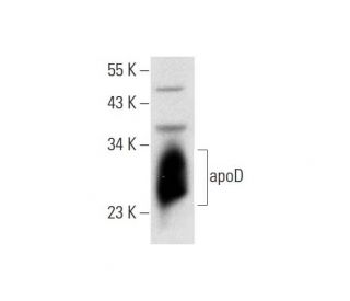
apoD Antibody (C-1): sc-373965
- apoD Antibody (C-1) is a mouse monoclonal IgG2b κ apoD antibody, cited in 4 publications, provided at 200 µg/ml
- raised against amino acids 1-189 representing full length apoD of human origin
- apoD Antibody (C-1) is recommended for detection of apoD of human origin by WB, IP, IF, IHC(P) and ELISA
- Anti-apoD Antibody (C-1) is available conjugated to agarose for IP; HRP for WB, IHC(P) and ELISA; and to either phycoerythrin or FITC for IF, IHC(P) and FCM
- also available conjugated to Alexa Fluor® 488, Alexa Fluor® 546, Alexa Fluor® 594 or Alexa Fluor® 647 for WB (RGB), IF, IHC(P) and FCM, and for use with RGB fluorescent imaging systems, such as iBright™ FL1000, FluorChem™, Typhoon, Azure and other comparable systems
- also available conjugated to Alexa Fluor® 680 or Alexa Fluor® 790 for WB (NIR), IF and FCM; for use with Near-Infrared (NIR) detection systems, such as LI-COR®Odyssey®, iBright™ FL1000, FluorChem™, Typhoon, Azure and other comparable systems
- m-IgG2b BP-HRP and m-IgGκ BP-HRP are the preferred secondary detection reagents for apoD Antibody (C-1) for WB and IHC(P) applications. These reagents are now offered in bundles with apoD Antibody (C-1) (see ordering information below).

apoD Antibody (C-1) is a mouse monoclonal IgG2b kappa light chain antibody that detects apoD protein of human origin by western blotting (WB), immunoprecipitation (IP), immunofluorescence (IF), immunohistochemistry with paraffin-embedded sections (IHCP), and enzyme-linked immunosorbent assay (ELISA). anti-apoD antibody (C-1) is available in both non-conjugated and various conjugated forms, including agarose, horseradish peroxidase (HRP), phycoerythrin (PE), fluorescein isothiocyanate (FITC), and multiple Alexa Fluor® conjugates. Apolipoprotein D (apoD) plays a crucial role in lipid transport and metabolism, functioning as a transporter protein that binds small hydrophobic molecules, such as arachidonic acid (AA). AA binding to apoD is significant as apoD participates in membrane phospholipid signal transduction pathways, which are essential for various cellular processes, including inflammation and cell signaling. apoD expression has been linked to cell cycle regulation and correlates with prognosis in several malignancies, including central nervous system astrocytomas and medulloblastomas, highlighting apoD′s potential as a biomarker in cancer research.
Alexa Fluor® is a trademark of Molecular Probes Inc., OR., USA
LI-COR® and Odyssey® are registered trademarks of LI-COR Biosciences
Ordering Information
| Product Name | Catalog # | UNIT | Price | Qty | FAVORITES | |
apoD Antibody (C-1) | sc-373965 | 200 µg/ml | $322.00 | |||
apoD Antibody (C-1): m-IgGκ BP-HRP Bundle | sc-522571 | 200 µg Ab, 40 µg BP | $361.00 | |||
apoD Antibody (C-1): m-IgG2b BP-HRP Bundle | sc-549042 | 200 µg Ab; 10 µg BP | $361.00 | |||
apoD Antibody (C-1) AC | sc-373965 AC | 500 µg/ml, 25% agarose | $424.00 | |||
apoD Antibody (C-1) HRP | sc-373965 HRP | 200 µg/ml | $322.00 | |||
apoD Antibody (C-1) FITC | sc-373965 FITC | 200 µg/ml | $336.00 | |||
apoD Antibody (C-1) PE | sc-373965 PE | 200 µg/ml | $349.00 | |||
apoD Antibody (C-1) Alexa Fluor® 488 | sc-373965 AF488 | 200 µg/ml | $364.00 | |||
apoD Antibody (C-1) Alexa Fluor® 546 | sc-373965 AF546 | 200 µg/ml | $364.00 | |||
apoD Antibody (C-1) Alexa Fluor® 594 | sc-373965 AF594 | 200 µg/ml | $364.00 | |||
apoD Antibody (C-1) Alexa Fluor® 647 | sc-373965 AF647 | 200 µg/ml | $364.00 | |||
apoD Antibody (C-1) Alexa Fluor® 680 | sc-373965 AF680 | 200 µg/ml | $364.00 | |||
apoD Antibody (C-1) Alexa Fluor® 790 | sc-373965 AF790 | 200 µg/ml | $364.00 |