")
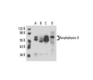
Amphiphysin II Antibody (G-10): sc-74487
- Amphiphysin II Antibody (G-10) is a mouse monoclonal IgG1 κ provided at 200 µg/ml
- raised against amino acids 421-520 mapping within an internal region of Amphiphysin II of human origin
- recommended for detection of all Amphiphysin II isoforms of mouse, rat and human origin by WB, IP, IF, IHC(P) and ELISA
- See Amphiphysin II (2F11): sc-23918 for Amphiphysin II antibody conjugates, including AC, HRP, FITC, PE, Alexa Fluor® 488, 594, 647, 680 and 790.
- m-IgG Fc BP-HRP is the preferred secondary detection reagent for Amphiphysin II Antibody (G-10) for WB and IHC(P) applications. This reagent is now offered in a bundle with Amphiphysin II Antibody (G-10) (see ordering information below).

QUICK LINKS
Amphiphysin II Antibody (G-10) is a mouse monoclonal IgG1 antibody that detects Amphiphysin II in mouse, rat, and human samples through various applications including western blotting (WB), immunoprecipitation (IP), immunofluorescence (IF), immunohistochemistry with paraffin-embedded sections (IHCP), and enzyme-linked immunosorbent assay (ELISA). Amphiphysin II is a crucial protein predominantly found in the brain, where Amphiphysin II plays a vital role in synaptic vesicle recycling, a process essential for neurotransmission. Amphiphysin II functions as a dimer and features a unique structure that includes a membrane-bending BAR domain, a central Clathrin and adaptor binding domain, and a C-terminal SH3 domain. The interaction of Amphiphysin II with the GTPase Dynamin via its SH3 domain is particularly important, as this interaction facilitates the binding of Dynamin to Clathrin-coated pits, leading to vesicle budding and efficient recycling of synaptic vesicles in neurons. Additionally, Amphiphysin II has a splice variant that is highly expressed in muscle tissue, which is involved in the formation and stabilization of the T tubule network, highlighting diverse functional roles across different tissues. Anti-Amphiphysin II antibody (G-10) is an invaluable tool for researchers studying synaptic function and membrane dynamics, providing insights into the molecular mechanisms underlying neurotransmission and muscle physiology.
Alexa Fluor® is a trademark of Molecular Probes Inc., OR., USA
LI-COR® and Odyssey® are registered trademarks of LI-COR Biosciences
Amphiphysin II Antibody (G-10) References:
- Bridging Integrator 1 (BIN1) Genotype Effects on Working Memory, Hippocampal Volume, and Functional Connectivity in Young Healthy Individuals. | Zhang, X., et al. 2015. Neuropsychopharmacology. 40: 1794-803. PMID: 25630570
- Inverse regulation of bridging integrator 1 and BCR-ABL1 in chronic myeloid leukemia. | Trino, S., et al. 2016. Tumour Biol. 37: 217-25. PMID: 26194865
- BIN1 regulates dynamic t-tubule membrane. | Fu, Y. and Hong, T. 2016. Biochim Biophys Acta. 1863: 1839-47. PMID: 26578114
- Post-Myocardial Infarction T-tubules Form Enlarged Branched Structures With Dysregulation of Junctophilin-2 and Bridging Integrator 1 (BIN-1). | Pinali, C., et al. 2017. J Am Heart Assoc. 6: PMID: 28473402
- Amphiphysin (BIN1) negatively regulates dynamin 2 for normal muscle maturation. | Cowling, BS., et al. 2017. J Clin Invest. 127: 4477-4487. PMID: 29130937
- Bridging Integrator-1 protein loss in Alzheimer's disease promotes synaptic tau accumulation and disrupts tau release. | Glennon, EB., et al. 2020. Brain Commun. 2: PMID: 32500121
- MTM1 overexpression prevents and reverts BIN1-related centronuclear myopathy. | Giraud, Q., et al. 2023. Brain. 146: 4158-4173. PMID: 37490306
- BIN1K358R suppresses glial response to plaques in mouse model of Alzheimer's disease. | Garcia-Agudo, LF., et al. 2024. Alzheimers Dement. 20: 2922-2942. PMID: 38460121
- The Alzheimer's disease risk gene BIN1 regulates activity-dependent gene expression in human-induced glutamatergic neurons. | Saha, O., et al. 2024. Mol Psychiatry. 29: 2634-2646. PMID: 38514804
- sTREM2 Mediates the Correlation Between BIN1 Gene Polymorphism and Tau Pathology in Alzheimer's Disease. | Guo, F., et al. 2024. J Alzheimers Dis. 101: 693-704. PMID: 39240638
Ordering Information
| Product Name | Catalog # | UNIT | Price | Qty | FAVORITES | |
Amphiphysin II Antibody (G-10) | sc-74487 | 200 µg/ml | $322.00 | |||
Amphiphysin II Antibody (G-10): m-IgG Fc BP-HRP Bundle | sc-539528 | 200 µg Ab; 10 µg BP | $361.00 |