Date published: 2026-4-4

1-800-457-3801

SCBT Portrait Logo
Seach Input

Amphiphysin I Antibody (C-1): sc-376397

5.0(4)
Write a reviewAsk a question

Datasheets
  • Amphiphysin I Antibody (C-1) is a mouse monoclonal IgG1 κ provided at 200 µg/ml
  • specific for an epitope mapping between amino acids 2-20 at the N-terminus of Amphiphysin of human origin
  • recommended for detection of Amphiphysin I of mouse, rat and human origin by WB, IP, IF and ELISA
  • m-IgG Fc BP-HRP and m-IgG1 BP-HRP are the preferred secondary detection reagents for Amphiphysin I Antibody (C-1) for WB applications. These reagents are now offered in bundles with Amphiphysin I Antibody (C-1) (see ordering information below).
    Gene Editing Promo Banner

    QUICK LINKS

    SEE ALSO...

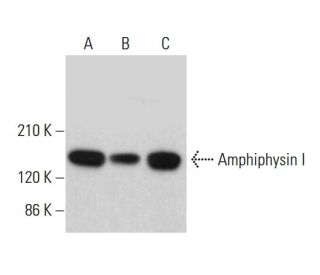
    Amphiphysin I Antibody (C-1) is a mouse monoclonal IgG1 antibody that detects Amphiphysin I in mouse, rat, and human samples through applications such as western blotting (WB), immunoprecipitation (IP), immunofluorescence (IF), and enzyme-linked immunosorbent assay (ELISA). Amphiphysin I is a crucial brain-enriched protein that plays a significant role in synaptic vesicle recycling, a process vital for neurotransmission. Anti-Amphiphysin I antibody (C-1) functions as a dimer and features a unique structure that includes a membrane-bending BAR domain, a central Clathrin and adaptor binding domain, and a C-terminal SH3 domain. The interaction between Amphiphysin I and the GTPase Dynamin, mediated by the SH3 domain, is essential for the proper binding of Dynamin to Clathrin-coated pits, facilitating vesicle budding and release at synapses. In addition to its role in the brain, Amphiphysin I is also implicated in membrane bending and curvature stabilization in various tissues, highlighting its importance in cellular dynamics. Notably, the mammalian Amphiphysins, including Amphiphysin I and its splice variant Amphiphysin II, share a similar overall structure, yet they exhibit distinct functional roles, particularly in muscle tissue where a splice form of Amphiphysin II contributes to the formation and stabilization of the T tubule network. Amphiphysin I monoclonal antibody (C-1) is an invaluable tool for researchers studying synaptic function and membrane dynamics across different biological systems.

    For Research Use Only. Not Intended for Diagnostic or Therapeutic Use.

    Ordering Information

    Product NameCatalog #UNITPriceQtyFAVORITES

    Amphiphysin I Antibody (C-1)

    sc-376397
    200 µg/ml
    $322.00

    Amphiphysin I Antibody (C-1): m-IgG Fc BP-HRP Bundle

    sc-540565
    200 µg Ab; 10 µg BP
    $361.00

    Amphiphysin I Antibody (C-1): m-IgG1 BP-HRP Bundle

    sc-542210
    200 µg Ab; 20 µg BP
    $361.00

    Amphiphysin I (C-1) Neutralizing Peptide

    sc-376397 P
    100 µg/0.5 ml
    $69.00

    What application is the blocking peptide sc-376397 P appropriate for?

    Asked by: Randy McDonald
    Thank you for your question. The blocking peptide is intended for use as a negative control, by pre-adsorbing the mouse monoclonal antibody against the antigen. For full protocol details, please contact our Technical Services Department or view our online protocol here: https://www.scbt.com/scbt/resources/protocols/peptide-neutralization
    Answered by: Technical Support
    Date published: 2017-02-25
    • y_2026, m_3, d_26, h_7CST
    • bvseo_bulk, prod_bvqa, vn_bulk_3.0.43
    • cp_1, bvpage1
    • co_hasquestionsanswers, tq_1
    • loc_en_US, sid_376397, prod, sort_[SortEntry(order=LAST_APPROVED_ANSWER_SUBMISSION_TIME, direction=DESCENDING)]
    • clientName_scbt
    • bvseo_sdk, java_sdk, bvseo-4.0.0
    • CLOUD, getContent, 102ms
    • QUESTIONS, PRODUCT
    Rated 5 out of 5 by from Strong bands in western blot analysis ofStrong bands in western blot analysis of Amphiphysin I expression in mouse brain, rat brain and human hippocampus tissue extracts. -SCBT QC
    Date published: 2015-07-10
    Rated 5 out of 5 by from Worked well with transfected lysate by WBWorked well with transfected lysate by WB. -SCBT QC
    Date published: 2015-06-17
    Rated 5 out of 5 by from Produced great immunofluorescence cytoplasmicProduced great immunofluorescence cytoplasmic staining in methanol-fixed HeLa cells. -SCBT QC
    Date published: 2015-03-15
    Rated 5 out of 5 by from Works well for western blotting; clean bandsWorks well for western blotting; clean bands in human hippocampus tissue extract and IMR-32 whole cell lysate. -SCBT QC
    Date published: 2015-03-08
    • y_2026, m_3, d_26, h_7
    • bvseo_bulk, prod_bvrr, vn_bulk_3.0.43
    • cp_1, bvpage1
    • co_hasreviews, tv_0, tr_4
    • loc_en_US, sid_376397, prod, sort_[SortEntry(order=SUBMISSION_TIME, direction=DESCENDING)]
    • clientName_scbt
    • bvseo_sdk, java_sdk, bvseo-4.0.0
    • CLOUD, getReviews, 10ms
    • REVIEWS, PRODUCT
    Amphiphysin I Antibody (C-1) is rated 5.0 out of 5 by 4.
    • y_2026, m_3, d_26, h_7
    • bvseo_bulk, prod_bvrr, vn_bulk_3.0.43
    • cp_1, bvpage1
    • co_hasreviews, tv_0, tr_4
    • loc_en_US, sid_376397, prod, sort_[SortEntry(order=SUBMISSION_TIME, direction=DESCENDING)]
    • clientName_scbt
    • bvseo_sdk, java_sdk, bvseo-4.0.0
    • CLOUD, getAggregateRating, 98ms
    • REVIEWS, PRODUCT