")
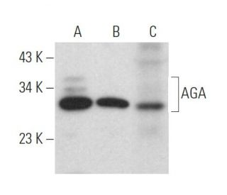
: sc-514075. Western blot analysis of AGA expression in Jurkat (A) and ALL-SIL (B) whole cell lysates and rat lung tissue extract (C).")
: sc-514075. Western blot analysis of AGA expression in non-transfected 293T: sc-117752 (A) and human AGA transfected 293T: sc-112982 (B) whole cell lysates and human heart tissue extract (C).")
AGA Antibody (H-8): sc-514075
- AGA Antibody (H-8) is a mouse monoclonal IgG1 κ AGA antibody provided at 200 µg/ml
- specific for an epitope mapping between amino acids 160-181 within an internal region of AGA of human origin
- AGA Antibody (H-8) is recommended for detection of AGA precursor and α chain of mouse, rat and human origin by WB, IP, IF and ELISA
- Anti-AGA Antibody (H-8) is available conjugated to agarose for IP; HRP for WB, IHC(P) and ELISA; and to either phycoerythrin or FITC for IF, IHC(P) and FCM
- also available conjugated to Alexa Fluor® 488, Alexa Fluor® 546, Alexa Fluor® 594 or Alexa Fluor® 647 for WB (RGB), IF, IHC(P) and FCM, and for use with RGB fluorescent imaging systems, such as iBright™ FL1000, FluorChem™, Typhoon, Azure and other comparable systems
- also available conjugated to Alexa Fluor® 680 or Alexa Fluor® 790 for WB (NIR), IF and FCM; for use with Near-Infrared (NIR) detection systems, such as LI-COR®Odyssey®, iBright™ FL1000, FluorChem™, Typhoon, Azure and other comparable systems
- m-IgG Fc BP-HRP and m-IgGκ BP-HRP are the preferred secondary detection reagents for AGA Antibody (H-8) for WB applications. These reagents are now offered in bundles with AGA Antibody (H-8) (see ordering information below).

QUICK LINKS
SEE ALSO...
AGA Antibody (H-8) is a mouse monoclonal IgG1 kappa light chain antibody that detects AGA protein of mouse, rat, and human origin by western blotting (WB), immunoprecipitation (IP), immunofluorescence (IF), and enzyme-linked immunosorbent assay (ELISA). Anti-AGA antibody (H-8) is available in both non-conjugated and various conjugated forms, including agarose, horseradish peroxidase (HRP), phycoerythrin (PE), fluorescein isothiocyanate (FITC), and multiple Alexa Fluor® conjugates. AGA protein, or aspartylglucosaminidase, is a 346 amino acid precursor that belongs to the Ntn-hydrolase family and is cleaved into an alpha chain and a beta chain. AGA protein is localized to the lysosome, where AGA protein plays a crucial role as a heterotetramer composed of two alpha and two beta chains, facilitating the cleavage of the GlcNAc-Asn bond that links oligosaccharides to target glycoproteins. Proper functioning of AGA protein is vital for the degradation of glycoproteins, and defects in the gene encoding AGA lead to aspartylglucosaminuria (AGU), a lysosomal storage disorder characterized by severe cognitive impairment and mild connective tissue abnormalities. The gene for AGA is located on human chromosome 4, which is notable for encoding nearly 6% of the human genome and containing some of the largest gene deserts among human chromosomes, highlighting the complexity and significance of genetic regulation in lysosomal function.
Alexa Fluor® is a trademark of Molecular Probes Inc., OR., USA
LI-COR® and Odyssey® are registered trademarks of LI-COR Biosciences
AGA Antibody (H-8) References:
- Molecular pathogenesis of a disease: structural consequences of aspartylglucosaminuria mutations. | Saarela, J., et al. 2001. Hum Mol Genet. 10: 983-95. PMID: 11309371
- Autoproteolytic activation of human aspartylglucosaminidase. | Saarela, J., et al. 2004. Biochem J. 378: 363-71. PMID: 14616088
- A novel aspartylglucosaminuria mutation affects translocation of aspartylglucosaminidase. | Saarela, J., et al. 2004. Hum Mutat. 24: 350-1. PMID: 15365992
- Elevation of plasma aspartylglucosaminidase is a useful marker for the congenital disorders of glycosylation type I (CDG I). | Jackson, M., et al. 2005. J Inherit Metab Dis. 28: 1197-8. PMID: 16435229
- Structural basis of aspartylglucosaminuria. | Saito, S., et al. 2008. Biochem Biophys Res Commun. 377: 1168-72. PMID: 18992224
- Plasma lysosomal enzyme activities in congenital disorders of glycosylation, galactosemia and fructosemia. | Michelakakis, H., et al. 2009. Clin Chim Acta. 401: 81-3. PMID: 19100247
- Human leucocyte glycosylasparaginase is an alpha/beta-heterodimer of 19 kDa alpha-subunit and 17 and 18 kDa beta-subunit. | Tollersrud, OK., et al. 1994. Biochem J. 300 (Pt 2): 541-4. PMID: 8002961
- Aspartylglycosaminuria: protein chemistry and molecular biology of the most common lysosomal storage disorder of glycoprotein degradation. | Mononen, I., et al. 1993. FASEB J. 7: 1247-56. PMID: 8405810
Ordering Information
| Product Name | Catalog # | UNIT | Price | Qty | FAVORITES | |
AGA Antibody (H-8) | sc-514075 | 200 µg/ml | $322.00 | |||
AGA Antibody (H-8): m-IgG Fc BP-HRP Bundle | sc-531020 | 200 µg Ab; 10 µg BP | $361.00 | |||
AGA Antibody (H-8): m-IgGκ BP-HRP Bundle | sc-524595 | 200 µg Ab, 40 µg BP | $361.00 | |||
AGA Antibody (H-8) AC | sc-514075 AC | 500 µg/ml, 25% agarose | $424.00 | |||
AGA Antibody (H-8) HRP | sc-514075 HRP | 200 µg/ml | $322.00 | |||
AGA Antibody (H-8) FITC | sc-514075 FITC | 200 µg/ml | $336.00 | |||
AGA Antibody (H-8) PE | sc-514075 PE | 200 µg/ml | $349.00 | |||
AGA Antibody (H-8) Alexa Fluor® 488 | sc-514075 AF488 | 200 µg/ml | $364.00 | |||
AGA Antibody (H-8) Alexa Fluor® 546 | sc-514075 AF546 | 200 µg/ml | $364.00 | |||
AGA Antibody (H-8) Alexa Fluor® 594 | sc-514075 AF594 | 200 µg/ml | $364.00 | |||
AGA Antibody (H-8) Alexa Fluor® 647 | sc-514075 AF647 | 200 µg/ml | $364.00 | |||
AGA Antibody (H-8) Alexa Fluor® 680 | sc-514075 AF680 | 200 µg/ml | $364.00 | |||
AGA Antibody (H-8) Alexa Fluor® 790 | sc-514075 AF790 | 200 µg/ml | $364.00 | |||
AGA (H-8) Neutralizing Peptide | sc-514075 P | 100 µg/0.5 ml | $69.00 |