")
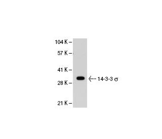
14-3-3 σ Antibody (5D7): sc-100638
- 14-3-3 σ Antibody (5D7) is a mouse monoclonal IgG1 κ, cited in 30 publications, provided at 100 µg/ml
- raised against recombinant 14-3-3 σ of human origin
- recommended for detection of 14-3-3 σ of human origin by WB, IP, IF, IHC(P) and ELISA
- See pan 14-3-3 (B-8): sc-133233 for 14-3-3 σ antibody conjugates, including AC, HRP, FITC, PE, Alexa Fluor® 488, 594, 647, 680 and 790.
- m-IgG Fc BP-HRP and m-IgG1 BP-HRP are the preferred secondary detection reagents for 14-3-3 σ Antibody (5D7) for WB and IHC(P) applications. These reagents are now offered in bundles with 14-3-3 σ Antibody (5D7) (see ordering information below).

QUICK LINKS
SEE ALSO...
14-3-3 σ Antibody (5D7) is a mouse monoclonal IgG1 kappa light chain antibody that detects 14-3-3 σ of human origin by western blotting (WB), immunoprecipitation (IP), immunofluorescence (IF), immunohistochemistry with paraffin-embedded sections (IHCP), and enzyme-linked immunosorbent assay (ELISA). Anti-14-3-3 σ antibody (5D7) is available in a non-conjugated form, providing flexibility for various experimental needs. 14-3-3 σ, also known as SFN, stratifin, HME1, or YWHAS, plays a crucial role in cellular signaling and regulation, particularly in cancer biology. 14-3-3 σ is predominantly expressed in stratified squamous keratinizing epithelium and functions as a secreted adaptor protein that interacts with various target proteins, including Keratin 17. 14-3-3 σ binding to Keratin 17 enhances the Akt/mTOR signaling pathway, promoting protein synthesis and cell growth, which are vital processes in tumor development and progression. 14-3-3 σ positively regulates tumor suppressor p53, influencing cell cycle checkpoints and apoptosis. This modulation of key pathways highlights 14-3-3 σ′s importance in maintaining cellular homeostasis and potential as a therapeutic target in cancer treatment. 14-3-3 σ monoclonal antibody (5D7) serves as an essential tool for researchers studying cancer biology and related fields.
Alexa Fluor® is a trademark of Molecular Probes Inc., OR., USA
LI-COR® and Odyssey® are registered trademarks of LI-COR Biosciences
14-3-3 σ Antibody (5D7) References:
- 14-3-3 sigma positively regulates p53 and suppresses tumor growth. | Yang, HY., et al. 2003. Mol Cell Biol. 23: 7096-107. PMID: 14517281
- A structural basis for 14-3-3sigma functional specificity. | Wilker, EW., et al. 2005. J Biol Chem. 280: 18891-8. PMID: 15731107
- 14-3-3sigma, a p53 regulator, suppresses tumor growth of nasopharyngeal carcinoma. | Yang, H., et al. 2006. Mol Cancer Ther. 5: 253-60. PMID: 16505098
- CpG island promoter methylation and silencing of 14-3-3sigma gene expression in LNCaP and Tramp-C1 prostate cancer cell lines is associated with methyl-CpG-binding protein MBD2. | Pulukuri, SM. and Rao, JS. 2006. Oncogene. 25: 4559-72. PMID: 16786000
- 14-3-3sigma controls mitotic translation to facilitate cytokinesis. | Wilker, EW., et al. 2007. Nature. 446: 329-32. PMID: 17361185
- 14-3-3 sigma isoform interacts with the cytoplasmic domain of the transmembrane BP180 in keratinocytes. | Li, Y., et al. 2007. J Cell Physiol. 212: 675-81. PMID: 17443672
- Methylation and intratumoural heterogeneity of 14-3-3 sigma in oral cancer. | Bhawal, UK., et al. 2007. Oncol Rep. 18: 817-24. PMID: 17786341
- Over-expression of 14-3-3sigma in budding colorectal cancer cells modulates cell migration in the presence of tenascin-C. | Ide, M., et al. 2007. Oncol Rep. 18: 1451-6. PMID: 17982629
- DNA methylation patterns of the CDH1, RARB, and SFN genes in choroid plexus tumors. | Losi-Guembarovski, R., et al. 2007. Cancer Genet Cytogenet. 179: 140-5. PMID: 18036402
- Detection of 14-3-3 sigma (σ) promoter methylation as a noninvasive biomarker using blood samples for breast cancer diagnosis. | Ye, M., et al. 2017. Oncotarget. 8: 9230-9242. PMID: 27999208
- 14-3-3σ and Its Modulators in Cancer. | Aljabal, G. and Yap, BK. 2020. Pharmaceuticals (Basel). 13: PMID: 33287252
- 14-3-3σ-NEDD4L axis promotes ubiquitination and degradation of HIF-1α in colorectal cancer. | Liu, S., et al. 2023. Cell Rep. 42: 112870. PMID: 37494179
Ordering Information
| Product Name | Catalog # | UNIT | Price | Qty | FAVORITES | |
14-3-3 σ Antibody (5D7) | sc-100638 | 100 µg/ml | $339.00 | |||
14-3-3 σ Antibody (5D7): m-IgG Fc BP-HRP Bundle | sc-539703 | 100 µg Ab; 10 µg BP | $361.00 | |||
14-3-3 σ Antibody (5D7): m-IgG1 BP-HRP Bundle | sc-541661 | 100 µg Ab; 20 µg BP | $361.00 |