
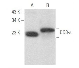
CD3-epsilon Antibody (G-5): sc-137096
- CD3-ε Antibody (G-5) is a mouse monoclonal IgG1 κ, cited in 1 publications, provided at 200 µg/ml
- raised against amino acids 1-207 representing full length CD3-ε of human origin
- recommended for detection of CD3-ε of mouse, rat and human origin by WB, IP, IF, IHC(P) and ELISA
- See CD3-ε (UCH-T1): sc-1179 for CD3-ε antibody conjugates, including AC, HRP, FITC, PE, Alexa Fluor® 488, 594, 647, 680 and 790.
- m-IgG Fc BP-HRP, m-IgG1 BP-HRP and m-IgGκ BP-HRP are the preferred secondary detection reagents for CD3-ε Antibody (G-5) for WB and IHC(P) applications. These reagents are now offered in bundles with CD3-ε Antibody (G-5) (see ordering information below).

CD3-ε Antibody (G-5) is a mouse monoclonal IgG1 antibody that detects CD3-ε in mouse, rat, and human samples through various applications including western blotting (WB), immunoprecipitation (IP), immunofluorescence (IF), immunohistochemistry, and enzyme-linked immunosorbent assay (ELISA). CD3-ε plays a crucial role in the immune response as a component of the T cell receptor (TCR) complex, which is essential for T cell activation and signaling. Located on the surface of T cells, CD3-ε is part of a multisubunit complex that transmits signals upon recognition of foreign antigens, facilitating the transition of T cells from a resting state to an activated state. CD3-ε, along with associated chains, ensures T cells can effectively respond to pathogens. CD3-ε contains an immunoreceptor tyrosine-based activation motif (ITAM) that is critical for initiating downstream signaling cascades, making CD3-ε a key player in the regulation of T cell responses. Anti-CD3-ε antibody (G-5) serves as an invaluable tool for researchers studying T cell biology and the mechanisms of immune activation, providing insights into both normal immune function and potential therapeutic targets in immunological diseases.
Alexa Fluor® is a trademark of Molecular Probes Inc., OR., USA
LI-COR® and Odyssey® are registered trademarks of LI-COR Biosciences
Contains Mercury: All HRP conjugates (including bundle forms) contain thiomersal, a mercury compound. Do not dispose in trash. Dispose as hazardous chemical waste according to local, state and federal regulations.
Download thiomersal SDS
CD3-epsilon Antibody (G-5) References:
- Ontogeny of human natural killer (NK) cells: fetal NK cells mediate cytolytic function and express cytoplasmic CD3 epsilon,delta proteins. | Phillips, JH., et al. 1992. J Exp Med. 175: 1055-66. PMID: 1372642
- Activation of T cells by a tyrosine kinase activation domain in the cytoplasmic tail of CD3 epsilon. | Letourneur, F. and Klausner, RD. 1992. Science. 255: 79-82. PMID: 1532456
- Interaction between the N-terminal SH3 domain of Nck-alpha and CD3-epsilon-derived peptides: non-canonical and canonical recognition motifs. | Santiveri, CM., et al. 2009. Biochim Biophys Acta. 1794: 110-7. PMID: 18955169
- Regulation of thymocyte development through CD3. II. Expression of T cell receptor beta CD3 epsilon and maturation to the CD4+8+ stage are highly correlated in individual thymocytes. | Levelt, CN., et al. 1993. J Exp Med. 178: 1867-75. PMID: 7504052
- Normal development and function of natural killer cells in CD3 epsilon delta 5/delta 5 mutant mice. | Renard, V., et al. 1995. Proc Natl Acad Sci U S A. 92: 7545-9. PMID: 7638228
- The T cell receptor associated CD3-epsilon protein is phosphorylated upon T cell activation in the two tyrosine residues of a conserved signal transduction motif. | Sancho, J., et al. 1993. Eur J Immunol. 23: 1636-42. PMID: 7686857
- Frequent expression of CD3 epsilon in CD3 (Leu 4)-negative nasal T-cell lymphomas. | Ohno, T., et al. 1995. Leukemia. 9: 44-52. PMID: 7845028
- Reliable method for the simultaneous detection of cytoplasmic and surface CD3 epsilon expression by murine lymphoid cells. | Franek, KJ., et al. 1994. Cytometry. 17: 224-36. PMID: 7851158
- Restoration of early thymocyte differentiation in T-cell receptor beta-chain-deficient mutant mice by transmembrane signaling through CD3 epsilon. | Levelt, CN., et al. 1993. Proc Natl Acad Sci U S A. 90: 11401-5. PMID: 8248261
- Tyrosine phosphorylation of the CD3-epsilon subunit of the T cell antigen receptor mediates enhanced association with phosphatidylinositol 3-kinase in Jurkat T cells. | de Aós, I., et al. 1997. J Biol Chem. 272: 25310-8. PMID: 9312149
Ordering Information
| Product Name | Catalog # | UNIT | Price | Qty | FAVORITES | |
CD3-ε Antibody (G-5) | sc-137096 | 200 µg/ml | $322.00 | |||
CD3-ε Antibody (G-5): m-IgG Fc BP-HRP Bundle | sc-553218 | 200 µg Ab; 10 µg BP | $361.00 | |||
CD3-ε Antibody (G-5): m-IgGκ BP-HRP Bundle | sc-553217 | 200 µg Ab; 40 µg BP | $361.00 | |||
CD3-ε Antibody (G-5): m-IgG1 BP-HRP Bundle | sc-553219 | 200 µg Ab; 20 µg BP | $361.00 |