Date published: 2026-6-2

1-800-457-3801

SCBT Portrait Logo
Seach Input

BUB1 Antibody (B-3): sc-365685

4.2(6)
Write a reviewAsk a question

Datasheets
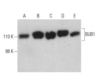
  • BUB1 Antibody (B-3) is a mouse monoclonal IgG3 κ BUB1 antibody, cited in 10 publications, provided at 200 µg/ml
  • raised against amino acids 786-1085 mapping at the C-terminus of BUB1 of human origin
  • recommended for detection of BUB1 of mouse, rat and human origin by WB, IP, IF, IHC(P) and ELISA
  • m-IgG3 BP-HRP and m-IgGκ BP-HRP are the preferred secondary detection reagents for BUB1 Antibody (B-3) for WB and IHC(P) applications. These reagents are now offered in bundles with BUB1 Antibody (B-3) (see ordering information below).
Gene Editing Promo Banner

QUICK LINKS

SEE ALSO...

BUB1 Antibody (B-3) is a mouse monoclonal IgG3 kappa light chain antibody that detects BUB1 protein of mouse, rat, and human origin by western blotting (WB), immunoprecipitation (IP), immunofluorescence (IF), immunohistochemistry, and enzyme-linked immunosorbent assay (ELISA). Anti-BUB1 antibody (B-3) is available as the non-conjugated form. BUB1 protein plays a critical role in the mitotic checkpoint, a crucial mechanism that ensures proper chromosome segregation during cell division. BUB1 protein is located at the kinetochores, the protein structures on chromosomes where spindle fibers attach during mitosis. Such localization allows BUB1 protein to monitor microtubule attachment to kinetochores, ensuring chromosomes are correctly aligned before cell proceeds to anaphase. When BUB1 protein detects misalignment or improper attachment, a checkpoint response activates to halt the cell cycle, preventing potential genomic instability and aneuploidy. BUB1 protein′s role in maintaining genomic integrity highlights potential applications in cancer research, where mitotic checkpoint dysregulation can lead to tumorigenesis.

For Research Use Only. Not Intended for Diagnostic or Therapeutic Use.

Alexa Fluor® is a trademark of Molecular Probes Inc., OR., USA

LI-COR® and Odyssey® are registered trademarks of LI-COR Biosciences

BUB1 Antibody (B-3) References:

  1. Human BUBR1 is a mitotic checkpoint kinase that monitors CENP-E functions at kinetochores and binds the cyclosome/APC.  |  Chan, GK., et al. 1999. J Cell Biol. 146: 941-54. PMID: 10477750
  2. BUBR1 phosphorylation is regulated during mitotic checkpoint activation.  |  Li, W., et al. 1999. Cell Growth Differ. 10: 769-75. PMID: 10593653
  3. Bub3 gene disruption in mice reveals essential mitotic spindle checkpoint function during early embryogenesis.  |  Kalitsis, P., et al. 2000. Genes Dev. 14: 2277-82. PMID: 10995385
  4. CENP-E as an essential component of the mitotic checkpoint in vitro.  |  Abrieu, A., et al. 2000. Cell. 102: 817-26. PMID: 11030625
  5. Identification of a novel gene--SSK1--in human endothelial cells exposed to shear stress.  |  Donadelli, R., et al. 1998. Biochem Biophys Res Commun. 246: 881-7. PMID: 9618306
  6. The hBUB1 and hBUBR1 kinases sequentially assemble onto kinetochores during prophase with hBUBR1 concentrating at the kinetochore plates in mitosis.  |  Jablonski, SA., et al. 1998. Chromosoma. 107: 386-96. PMID: 9914370

Ordering Information

Product NameCatalog #UNITPriceQtyFAVORITES

BUB1 Antibody (B-3)

sc-365685
200 µg/ml
$322.00

BUB1 Antibody (B-3): m-IgGκ BP-HRP Bundle

sc-522328
200 µg Ab, 40 µg BP
$361.00

BUB1 Antibody (B-3): m-IgG3 BP-HRP Bundle

sc-550344
200 µg Ab; 40 µg BP
$361.00

Why does your datasheet say that the molecular weight of BUB1 is 150kDa but the Western blots show the protein being about 100kDa. What size band should I be looking for (Human protein)?

Asked by: tasutria
Thank you for your question. The listed molecular weight on the datasheet refers to a publication that cited the size for BUB1 as 150 kDa (PubMedID: 1142067), however published weights vary as low as 110 kDa for this protein (PubMedID: 10704439). The predicted weight based on sequence for BUB1 is ~122 kDa, and it undergoes post-translational modification including multiple phosphorylation sites, which changes it's observed weight on the gel. Our data falls within the observed range of 110-150 kDa. If you have any further questions please contact our Technical Services Department.
Answered by: Technical Service
Date published: 2017-03-01

I am using BUB1 (B-3): sc-365685 monoclonal antibody to stain paraffin embedded tissue. Do you recommend antigen retrieval when using this antibody?

Asked by: Germaine
We recommend performing antigen retrieval with sodium citrate buffer (pH 6) and heat. The full protocol can be found here: https://www.scbt.com/scbt/resources/protocols/immunoperoxidase-staining
Answered by: Technical Support
Date published: 2017-02-24
  • y_2026, m_6, d_1, h_13CST
  • bvseo_bulk, prod_bvqa, vn_bulk_3.0.46
  • cp_1, bvpage1
  • co_hasquestionsanswers, tq_2
  • loc_en_US, sid_365685, prod, sort_[SortEntry(order=LAST_APPROVED_ANSWER_SUBMISSION_TIME, direction=DESCENDING)]
  • clientName_scbt
  • bvseo_sdk, java_sdk, bvseo-4.0.0
  • CLOUD, getContent, 113ms
  • QUESTIONS, PRODUCT
Rated 1 out of 5 by from The antibody doesn't recognise BUB1.I did a siRNA experiment to check if the band that the antibody showed was Bub1 or not and the band didn't disappear. I double checked this with another antibody and the experiment worked properly so it has to be the antibody.
Date published: 2019-04-04
Rated 5 out of 5 by from Great antibody for IF!We received a sample and tested it in human colon carcinoma cells. It works really well in IF. Strong kinetochore signal only with PFA fixation!
Date published: 2017-11-09
Rated 5 out of 5 by from 该抗体对试验帮助很好The specificity of this antibody is very well and good at my research。
Date published: 2017-09-20
Rated 5 out of 5 by from Versatile AntibodyThis antibody is well-cited for a variety of applications including WB, IP, IF, and even IHC and ELISA. Strong, clear signal with little to no background. Beautiful tissue staining. Works well in a variety of species including mouse, rat, and human samples.
Date published: 2017-01-23
Rated 4 out of 5 by from Strong bands seen in WB using HeLa and KStrong bands seen in WB using HeLa and K-562 whole cell lysates. -SCBT QC
Date published: 2015-04-16
Rated 5 out of 5 by from Exceptional immunoperoxidase cytoplasmicExceptional immunoperoxidase cytoplasmic staining in formalin fixed, paraffin-embedded human oral mucosa tissue. -SCBT QC
Date published: 2015-01-24
  • y_2026, m_5, d_31, h_13
  • bvseo_bulk, prod_bvrr, vn_bulk_3.0.46
  • cp_1, bvpage1
  • co_hasreviews, tv_0, tr_6
  • loc_en_US, sid_365685, prod, sort_[SortEntry(order=SUBMISSION_TIME, direction=DESCENDING)]
  • clientName_scbt
  • bvseo_sdk, java_sdk, bvseo-4.0.0
  • CLOUD, getReviews, 10ms
  • REVIEWS, PRODUCT
BUB1 Antibody (B-3) is rated 4.2 out of 5 by 6.
  • y_2026, m_5, d_31, h_13
  • bvseo_bulk, prod_bvrr, vn_bulk_3.0.46
  • cp_1, bvpage1
  • co_hasreviews, tv_0, tr_6
  • loc_en_US, sid_365685, prod, sort_[SortEntry(order=SUBMISSION_TIME, direction=DESCENDING)]
  • clientName_scbt
  • bvseo_sdk, java_sdk, bvseo-4.0.0
  • CLOUD, getAggregateRating, 87ms
  • REVIEWS, PRODUCT