")
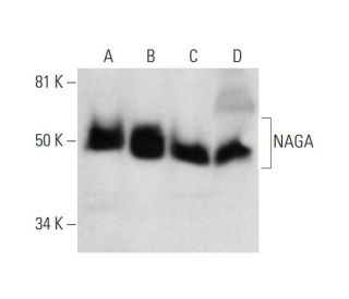
: sc-393485. Western blot analysis of NAGA expression in HeLa (A), JAR (B) and A-431 (C) whole cell lysates and human kidney tissue extract (D).")
: sc-393485. Western blot analysis of NAGA expression in HeLa (A), K-562 (B) and HCT-116 (C) whole cell lysates. Detection reagent used: m-IgG Fc BP-HRP: sc-525409.")
: sc-393485. Western blot analysis of NAGA expression in THP-1 whole cell lysate.")
NAGA Antibody (F-1): sc-393485
- NAGA Antibody (F-1) is a mouse monoclonal IgG1 κ NAGA antibody, cited in 1 publications, provided at 200 µg/ml
- raised against amino acids 335-411 mapping at the C-terminus of NAGA of human origin
- NAGA Antibody (F-1) is recommended for detection of NAGA of human origin by WB, IP, IF and ELISA
- Anti-NAGA Antibody (F-1) is available conjugated to agarose for IP; HRP for WB, IHC(P) and ELISA; and to either phycoerythrin or FITC for IF, IHC(P) and FCM
- also available conjugated to Alexa Fluor® 488, Alexa Fluor® 546, Alexa Fluor® 594 or Alexa Fluor® 647 for WB (RGB), IF, IHC(P) and FCM, and for use with RGB fluorescent imaging systems, such as iBright™ FL1000, FluorChem™, Typhoon, Azure and other comparable systems
- also available conjugated to Alexa Fluor® 680 or Alexa Fluor® 790 for WB (NIR), IF and FCM; for use with Near-Infrared (NIR) detection systems, such as LI-COR®Odyssey®, iBright™ FL1000, FluorChem™, Typhoon, Azure and other comparable systems
- m-IgG Fc BP-HRP, m-IgG1 BP-HRP and m-IgGκ BP-HRP are the preferred secondary detection reagents for NAGA Antibody (F-1) for WB applications. These reagents are now offered in bundles with NAGA Antibody (F-1) (see ordering information below).

QUICK LINKS
SEE ALSO...
NAGA Antibody (F-1) is a mouse monoclonal IgG1 kappa light chain antibody that detects NAGA protein of human origin by western blotting (WB), immunoprecipitation (IP), immunofluorescence (IF), and enzyme-linked immunosorbent assay (ELISA). anti-NAGA antibody (F-1) is available in both non-conjugated and various conjugated forms, including agarose, horseradish peroxidase (HRP), phycoerythrin (PE), fluorescein isothiocyanate (FITC), and multiple Alexa Fluor® conjugates. NAGA protein, also known as alpha-galactosidase B or GALB, is a 411 amino acid lysosomal enzyme that plays a vital role in the breakdown of glycolipids, which are essential for cellular membrane integrity and signaling. NAGA is located primarily in lysosomes, where NAGA functions to cleave alpha-N-acetylgalactosaminyl groups from glycoconjugates, facilitating the degradation of complex carbohydrates. Proper functioning of NAGA is crucial, as defects in this enzyme can lead to the accumulation of glycosphingolipids, resulting in severe neurological disorders such as Schindler disease, which presents with varying degrees of intellectual impairment and other systemic manifestations. Mapping to human chromosome 22q13, mutations in the NAGA gene are responsible for this autosomal recessive disorder, highlighting NAGA′s importance in maintaining cellular homeostasis and the potential consequences of dysfunction.
Alexa Fluor® is a trademark of Molecular Probes Inc., OR., USA
LI-COR® and Odyssey® are registered trademarks of LI-COR Biosciences
NAGA Antibody (F-1) References:
- A new case of alpha-N-acetylgalactosaminidase deficiency with angiokeratoma corporis diffusum, with Ménière's syndrome and without mental retardation. | Kodama, K., et al. 2001. Br J Dermatol. 144: 363-8. PMID: 11251574
- Human alpha-N-acetylgalactosaminidase (alpha-NAGA) deficiency: no association with neuroaxonal dystrophy? | Bakker, HD., et al. 2001. Eur J Hum Genet. 9: 91-6. PMID: 11313741
- Localization of a gene for human alpha-galactosidase B (= n-acetyl-alpha-d-galactosaminidase) on chromosome 22. | de Groot, PG., et al. 1978. Hum Genet. 44: 305-12. PMID: 215508
- Schindler disease: the molecular lesion in the alpha-N-acetylgalactosaminidase gene that causes an infantile neuroaxonal dystrophy. | Wang, AM., et al. 1990. J Clin Invest. 86: 1752-6. PMID: 2243144
- Regional localization of the genes coding for human ACO2, ARSA, and NAGA on chromosome 22. | Geurts van Kessel, AH., et al. 1980. Cytogenet Cell Genet. 28: 169-72. PMID: 7192199
- alpha-N-acetylgalactosaminidase deficiency with mild clinical manifestations and difficult biochemical diagnosis. | de Jong, J., et al. 1994. J Pediatr. 125: 385-91. PMID: 8071745
- Human alpha-N-acetylgalactosaminidase (alpha-NAGA) deficiency: new mutations and the paradox between genotype and phenotype. | Keulemans, JL., et al. 1996. J Med Genet. 33: 458-64. PMID: 8782044
Ordering Information
| Product Name | Catalog # | UNIT | Price | Qty | FAVORITES | |
NAGA Antibody (F-1) | sc-393485 | 200 µg/ml | $322.00 | |||
NAGA Antibody (F-1): m-IgG Fc BP-HRP Bundle | sc-530467 | 200 µg Ab; 10 µg BP | $361.00 | |||
NAGA Antibody (F-1): m-IgGκ BP-HRP Bundle | sc-523961 | 200 µg Ab, 40 µg BP | $361.00 | |||
NAGA Antibody (F-1): m-IgG1 BP-HRP Bundle | sc-544066 | 200 µg Ab; 20 µg BP | $361.00 | |||
NAGA Antibody (F-1) AC | sc-393485 AC | 500 µg/ml, 25% agarose | $424.00 | |||
NAGA Antibody (F-1) HRP | sc-393485 HRP | 200 µg/ml | $322.00 | |||
NAGA Antibody (F-1) FITC | sc-393485 FITC | 200 µg/ml | $336.00 | |||
NAGA Antibody (F-1) PE | sc-393485 PE | 200 µg/ml | $349.00 | |||
NAGA Antibody (F-1) Alexa Fluor® 488 | sc-393485 AF488 | 200 µg/ml | $364.00 | |||
NAGA Antibody (F-1) Alexa Fluor® 546 | sc-393485 AF546 | 200 µg/ml | $364.00 | |||
NAGA Antibody (F-1) Alexa Fluor® 594 | sc-393485 AF594 | 200 µg/ml | $364.00 | |||
NAGA Antibody (F-1) Alexa Fluor® 647 | sc-393485 AF647 | 200 µg/ml | $364.00 | |||
NAGA Antibody (F-1) Alexa Fluor® 680 | sc-393485 AF680 | 200 µg/ml | $364.00 | |||
NAGA Antibody (F-1) Alexa Fluor® 790 | sc-393485 AF790 | 200 µg/ml | $364.00 |