
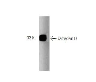

cathepsin D Antibody (49): sc-136282
- cathepsin D Antibody (49) is a mouse monoclonal IgG2a, cited in 4 publications, provided at 50 µg/0.5 ml
- recommended for detection of cathepsin D of human origin by WBIP; not recommended for IP
- See cathepsin D (D-7): sc-377299 for cathepsin D antibody conjugates, including AC, HRP, FITC, PE, Alexa Fluor® 488, 594, 647, 680 and 790.
- At present, we have not yet completed the identification of the preferred secondary detection reagent(s) for cathepsin D Antibody (49). This work is in progress.

QUICK LINKS
SEE ALSO...
cathepsin D Antibody (49) is a mouse monoclonal IgG2a antibody with validated applications in western blotting (WB) and immunoprecipitation (IP) and reactivity to human proteins. Cathepsin D, also referred to as CTSD, is an aspartyl protease that is predominantly located in the lysosomes, where CTSD plays a central role in protein degradation and turnover. Structurally, cathepsin D is synthesized as a precursor protein that undergoes proteolytic cleavage to form an active enzyme, consisting of a heavy and light chain linked by disulfide bonds. This structure is essential for CTSD stability and enzymatic function within the acidic lysosomal environment. In addition to degrading intracellular proteins, cathepsin D is involved in processing specific protein precursors to generate bioactive peptides that regulate processes such as apoptosis, cell growth, and immune responses. CTSD activity is regulated through structure and localization, with disruptions in these processes linked to pathological conditions, including cancer and neurodegenerative diseases. The gene encoding cathepsin D contains regulatory elements such as Sp1 and AP-2 binding sites, which control CTSD expression in response to cellular signals. Anti-cathepsin D antibody (49) is an effective tool for studying the structural and functional roles of cathepsin D in health and disease.
Alexa Fluor® is a trademark of Molecular Probes Inc., OR., USA
LI-COR® and Odyssey® are registered trademarks of LI-COR Biosciences
cathepsin D Antibody (49) References:
- Cathepsin D is up-regulated in inflammatory bowel disease macrophages. | Hausmann, M., et al. 2004. Clin Exp Immunol. 136: 157-67. PMID: 15030527
- Cathepsin D in the malignant progression of neoplastic diseases (review). | Leto, G., et al. 1992. Anticancer Res. 12: 235-40. PMID: 1567172
- Dual role of cathepsin D: ligand and protease. | Fusek, M. and Vetvicka, V. 2005. Biomed Pap Med Fac Univ Palacky Olomouc Czech Repub. 149: 43-50. PMID: 16170387
- Cathepsin D and prognosis in breast cancer. | Tandon, AK., et al. 1990. N Engl J Med. 322: 297-302. PMID: 2296271
- Overexpression of cathepsin D in malignant melanoma. | Zhu, L., et al. 2013. Fukuoka Igaku Zasshi. 104: 370-5. PMID: 24511668
- Cathepsin D in Podocytes Is Important in the Pathogenesis of Proteinuria and CKD. | Yamamoto-Nonaka, K., et al. 2016. J Am Soc Nephrol. 27: 2685-700. PMID: 26823550
- Effect of Curcumin on Serum Cathepsin D in Patients with Metabolic Syndrome. | Shirmohammadi, L., et al. 2020. Cardiovasc Hematol Disord Drug Targets. 20: 116-121. PMID: 31538907
- Myocardial cathepsin D is downregulated in sudden cardiac death. | Kakimoto, Y., et al. 2020. PLoS One. 15: e0230375. PMID: 32176724
- Identification of BAG2 and Cathepsin D as Plasma Biomarkers for Parkinson's Disease. | Kang, J., et al. 2021. Clin Transl Sci. 14: 606-616. PMID: 33202088
- Cathepsin D is membrane-associated in macrophage endosomes. | Diment, S., et al. 1988. J Biol Chem. 263: 6901-7. PMID: 3360812
Ordering Information
| Product Name | Catalog # | UNIT | Price | Qty | FAVORITES | |
cathepsin D Antibody (49) | sc-136282 | 50 µg/0.5 ml | $322.00 |