")
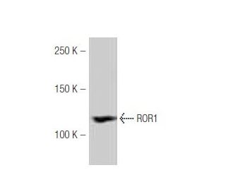
: sc-130386. Western blot analysis of ROR1 expression in Hep G2 whole cell lysate.")
ROR1 Antibody (60-D): sc-130386
- ROR1 Antibody (60-D) is a mouse monoclonal IgG1 κ ROR1 antibody, cited in 5 publications, provided at 100 µg/ml
- raised against an internal region of ROR1 of human origin
- recommended for detection of ROR1 of mouse, rat and human origin by WB and IP
- m-IgG Fc BP-HRP, m-IgG1 BP-HRP and m-IgGκ BP-HRP are the preferred secondary detection reagents for ROR1 Antibody (60-D) for WB applications. These reagents are now offered in bundles with ROR1 Antibody (60-D) (see ordering information below).

QUICK LINKS
SEE ALSO...
ROR1 Antibody (60-D) is a mouse monoclonal IgG1 kappa light chain antibody that detects ROR1 protein of mouse, rat, and human origin by western blotting (WB) and immunoprecipitation (IP). Anti-ROR1 antibody (60-D) is available as a non-conjugated format. ROR1 plays a crucial role in organogenesis and neuronal differentiation, making ROR1 significant in developmental biology and potential therapeutic applications. Located primarily on the cell surface, ROR1 is involved in various signaling pathways that influence cell growth and differentiation. ROR1 expression is notably increased in certain hematological malignancies, such as acute lymphoblastic leukemia and chronic lymphocytic leukemia, highlighting ROR1 potential as a target for immunotherapy. The ROR family, which includes ROR1 and ROR2, is characterized by unique structural features, including intracellular tyrosine kinase domains and extracellular Frizzled-like cysteine-rich domains, which are essential for cellular signaling. The conservation of ROR1 across species, from C. elegans to mammals, underscores ROR1 fundamental role in biological processes. ROR1 monoclonal antibody (60-D) serves as a valuable tool for researchers investigating ROR1 implications in both normal physiology and disease states.
Alexa Fluor® is a trademark of Molecular Probes Inc., OR., USA
LI-COR® and Odyssey® are registered trademarks of LI-COR Biosciences
ROR1 Antibody (60-D) References:
- Expression and function of the Ror-family receptor tyrosine kinases during development: lessons from genetic analyses of nematodes, mice, and humans. | Yoda, A., et al. 2003. J Recept Signal Transduct Res. 23: 1-15. PMID: 12680586
- Expression and subcellular localization of Ror tyrosine kinase receptors are developmentally regulated in cultured hippocampal neurons. | Paganoni, S. and Ferreira, A. 2003. J Neurosci Res. 73: 429-40. PMID: 12898527
- A novel family of cell surface receptors with tyrosine kinase-like domain. | Masiakowski, P. and Carroll, RD. 1992. J Biol Chem. 267: 26181-90. PMID: 1334494
- Unique cell surface expression of receptor tyrosine kinase ROR1 in human B-cell chronic lymphocytic leukemia. | Baskar, S., et al. 2008. Clin Cancer Res. 14: 396-404. PMID: 18223214
- Overexpression of orphan receptor tyrosine kinase Ror1 as a putative tumor-associated antigen in Iranian patients with acute lymphoblastic leukemia. | Shabani, M., et al. 2007. Tumour Biol. 28: 318-26. PMID: 18354269
- Ror1, a cell surface receptor tyrosine kinase is expressed in chronic lymphocytic leukemia and may serve as a putative target for therapy. | Daneshmanesh, AH., et al. 2008. Int J Cancer. 123: 1190-5. PMID: 18546292
- Expression profile of orphan receptor tyrosine kinase (ROR1) and Wilms' tumor gene 1 (WT1) in different subsets of B-cell acute lymphoblastic leukemia. | Shabani, M., et al. 2008. Leuk Lymphoma. 49: 1360-7. PMID: 18604725
- Wnt-ligand-dependent interaction of TAK1 (TGF-beta-activated kinase-1) with the receptor tyrosine kinase Ror2 modulates canonical Wnt-signalling. | Winkel, A., et al. 2008. Cell Signal. 20: 2134-44. PMID: 18762249
- Tyrosine Kinase ROR1 as a Target for Anti-Cancer Therapies. | Zhao, Y., et al. 2021. Front Oncol. 11: 680834. PMID: 34123850
- The signaling pathways activated by ROR1 in cancer. | Quezada, MJ. and Lopez-Bergami, P. 2023. Cell Signal. 104: 110588. PMID: 36621728
- IGFBP5 is an ROR1 ligand promoting glioblastoma invasion via ROR1/HER2-CREB signaling axis. | Lin, W., et al. 2023. Nat Commun. 14: 1578. PMID: 36949068
- ROR1 for Lymphoid Cancers. | Schuster, SJ. 2022. NEJM Evid. 1: EVIDe2100014. PMID: 38319244
Ordering Information
| Product Name | Catalog # | UNIT | Price | Qty | FAVORITES | |
ROR1 Antibody (60-D) | sc-130386 | 100 µg/ml | $339.00 | |||
ROR1 Antibody (60-D): m-IgG Fc BP-HRP Bundle | sc-537328 | 100 µg Ab; 10 µg BP | $361.00 | |||
ROR1 Antibody (60-D): m-IgGκ BP-HRP Bundle | sc-534569 | 100 µg Ab; 40 µg BP | $361.00 | |||
ROR1 Antibody (60-D): m-IgG1 BP-HRP Bundle | sc-545139 | 100 µg Ab; 20 µg BP | $361.00 |