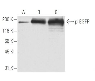
Date published: 2025-9-9

00800 4573 8000

SCBT Portrait Logo
Seach Input

p-EGFR Antikörper (9H2): sc-57545

4.8(8)
Produkt bewertenBitte stellen Sie eine Frage

Datenblätter
  • p-EGFR Antikörper 9H2 ist ein monoklonales IgG1 aus der Maus, verwendet in 12 wissenschaftlichen Veröffentlichungen, in einer Menge von 50 µg/0.5 ml
  • gegen ein EFGR-Phosphopeptid mit Ursprung human
  • Empfohlen für die Detektion von Tyr 1173 phosphorylated EGFR aus der Spezies mouse, rat, human und canine per WB, IP und IF; nicht kreuzreaktiv mit dem non-phosphorylated EGFR nor with unrelated Tyrosine-phosphorylated proteins
  • Als Direktkonjugat zur Detektion von p-EGFR wird EGFR (A-10): sc-373746 angeboten; Primärantikörper konjugiert mit AC, HRP, FITC, PE, Alexa Fluor® 488, 594, 647, 680 und 790.
  • Bitte kontaktieren Sie uns um ein 10 µg GRATIS Sample vom p-EGFR (9H2): sc-57545 zu erhalten.
  • m-IgG Fc BP-HRP und 1 BP-HRP">m-IgG1 BP-HRP sind die bevorzugten sekundären Nachweisreagenzien für p-EGFR Antikörper (9H2) for WB applications.. Diese Reagenzien werden jetzt in Bündeln mit p-EGFR Antikörper (9H2) angeboten(siehe Bestellinformationen unten).

Direktverknüpfungen

Siehe auch...

Der p-EGFR-Antikörper (9H2) ist ein monoklonaler IgG1-Antikörper der Maus, der den phosphorylierten EGFR Tyr 1173 in Proben von Mäusen, Ratten, Menschen und Hunden durch Anwendungen wie Western Blotting (WB), Immunopräzipitation (IP) und Immunfluoreszenz (IF) nachweist. Der p-EGFR-monoklonale Antikörper (9H2) ist in nicht konjugierter Form erhältlich, was vielseitige Versuchsaufbauten ermöglicht. Der epidermale Wachstumsfaktor-Rezeptor (EGFR) spielt eine entscheidende Rolle bei der Signalübertragung in der Zelle und vermittelt zelluläre Reaktionen auf Wachstumsfaktoren, die für Prozesse wie Zellproliferation, Differenzierung und Überleben von entscheidender Bedeutung sind. Der hauptsächlich auf der Zelloberfläche befindliche EGFR wird durch die Bindung an Liganden wie EGF oder TGFα aktiviert, was eine Kaskade intrazellulärer Signalwege auslöst, die für die normale Zellfunktion und -entwicklung unerlässlich sind. Die Phosphorylierung spezifischer Tyrosinreste, insbesondere Tyr 1173, ist für die Aktivierung des EGFR und die anschließende Signalübertragung von entscheidender Bedeutung, da die Phosphorylierung die Rekrutierung nachgeschalteter Signalproteine erleichtert, darunter die Phosphotyrosin-Bindungsdomäne von GRB2. Diese Interaktion ist für die Aktivierung des Ras-Signalwegs von entscheidender Bedeutung, der für das Zellwachstum und die Zellteilung von wesentlicher Bedeutung ist. Eine Fehlregulation der EGFR-Signalübertragung ist häufig an verschiedenen Krebsarten beteiligt, sodass der p-EGFR (9H2)-Antikörper ein wertvolles Instrument für die Erforschung der Krebsbiologie und therapeutischer Interventionen darstellt.

Ausschließlich für Forschungszwecke. Nicht für diagnostische oder therapeutische Zwecke bestimmt.

Bestellinformation

ProduktKatalog #EINHEITPreisANZAHLFavoriten

p-EGFR Antikörper (9H2)

sc-57545
50 µg/0.5 ml
$316.00

p-EGFR (9H2): m-IgG Fc BP-HRP Bundle

sc-539060
50 µg Ab; 10 µg BP
$354.00

p-EGFR (9H2): m-IgG1 BP-HRP Bundle

sc-541235
50 µg Ab; 20 µg BP
$354.00

How much protein do you recommend loading on the gel for Western blot detection using p-EGFR (9H2): sc-57545 monoclonal antibody?

Gefragt von: jenniferc
We recommend loading 40–60 µg whole cell lysate, 10–20 µg nuclear extract, 5-10 µg transfected lysate, or 10–20 ng purified protein per lane. A link to our complete Western blotting protocol is provided here for your reference. https://www.scbt.com/scbt/resources/protocols/western-immuno-blotting
Beantwortet von: Santa Cruz TS
Veröffentlichungsdatum: 2017-02-02
  • y_2025, m_9, d_6, h_5CST
  • bvseo_bulk, prod_bvqa, vn_bulk_3.0.42
  • cp_1, bvpage1
  • co_hasquestionsanswers, tq_1
  • loc_de_DE, sid_57545, prod, sort_[SortEntry(order=LAST_APPROVED_ANSWER_SUBMISSION_TIME, direction=DESCENDING)]
  • clientName_scbt
  • bvseo_sdk, java_sdk, bvseo-4.0.0
  • CLOUD, getContent, 119ms
  • QUESTIONS, PRODUCT
Rated 5 von 5 von aus very good antibodyI can detect band! The antibody was used for Western Blot in hepa1c1c7 cells, it works at a concentration of 1: 100 ~ 200.
Veröffentlichungsdatum: 2022-12-07
Rated 4 von 5 von aus GoodThe antibody was used for Western Blot in breast carcinoma cells, it works at a concentration of 1: 100.
Veröffentlichungsdatum: 2020-10-16
Rated 4 von 5 von aus Works wellused for WB looking at mouse tissue samples using 1:200. Provides a faint but clear band.
Veröffentlichungsdatum: 2018-03-22
Rated 5 von 5 von aus Great antibodyi got this antibody as free trial size and worked so well on mouse mammary tissue lysates.
Veröffentlichungsdatum: 2017-12-15
Rated 5 von 5 von aus Great phospho-detectionGreat, published antibody for p-EGFR detection recommended for use in mouse, rat, human, and canine. Strong bands in WB for phosphorylated samples.
Veröffentlichungsdatum: 2017-01-30
Rated 5 von 5 von aus Working great with dSTORM IF with HeLa andWorking great with dSTORM IF with HeLa and Cos7 cells.
Veröffentlichungsdatum: 2015-03-30
Rated 5 von 5 von aus Good Western blot analysis of EGFR phosphorylationGood Western blot analysis of EGFR phosphorylation in non-stimulated, EGF stimulated and pervanadate stimulated A549 whole cell lysates. - SCBT QC
Veröffentlichungsdatum: 2014-02-09
  • y_2025, m_9, d_6, h_5
  • bvseo_bulk, prod_bvrr, vn_bulk_3.0.42
  • cp_1, bvpage1
  • co_hasreviews, tv_1, tr_7
  • loc_de_DE, sid_57545, prod, sort_[SortEntry(order=SUBMISSION_TIME, direction=DESCENDING)]
  • clientName_scbt
  • bvseo_sdk, java_sdk, bvseo-4.0.0
  • CLOUD, getReviews, 19ms
  • REVIEWS, PRODUCT
p-EGFR Antikörper (9H2) wurde bewertet mit 4.8 von 5 von 8.
  • y_2025, m_9, d_6, h_5
  • bvseo_bulk, prod_bvrr, vn_bulk_3.0.42
  • cp_1, bvpage1
  • co_hasreviews, tv_1, tr_7
  • loc_de_DE, sid_57545, prod, sort_[SortEntry(order=SUBMISSION_TIME, direction=DESCENDING)]
  • clientName_scbt
  • bvseo_sdk, java_sdk, bvseo-4.0.0
  • CLOUD, getAggregateRating, 111ms
  • REVIEWS, PRODUCT