")
EGFR Antibody (528): sc-120
- EGFR Antibody (528) is a mouse monoclonal IgG2a κ, cited in 361 publications, provided at 200 µg/ml
- mapping to a cell surface epitope of EGFR of human origin
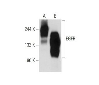
- Anti-EGFR Antibody (528) is recommended for detection of EGFR of mouse, rat and human origin by WB (non-reducing), IP, IF and FCM; also reactive with additional species, including canine
- EGFR Antibody (528) is available conjugated to agarose for IP; and to either phycoerythrin or FITC for IF, IHC(P) and FCM
- also available conjugated to Alexa Fluor® 488, Alexa Fluor® 546, Alexa Fluor® 594 or Alexa Fluor® 647 for WB (RGB), IF, IHC(P) and FCM, and for use with RGB fluorescent imaging systems, such as iBright™ FL1000, FluorChem™, Typhoon, Azure and other comparable systems
- also available conjugated to Alexa Fluor® 680 or Alexa Fluor® 790 for WB (NIR), IF and FCM; for use with Near-Infrared (NIR) detection systems, such as LI-COR®Odyssey®, iBright™ FL1000, FluorChem™, Typhoon, Azure and other comparable systems
- also available conjugated to biotin for WB, IHC(P) and ELISA
- m-IgG Fc BP-HRP and m-IgG2a BP-HRP are the preferred secondary detection reagents for EGFR Antibody (528). These reagents are now offered in bundles with EGFR Antibody (528) (see ordering information below).

QUICK LINKS
SEE ALSO...
EGFR Antibody (528) is a mouse monoclonal IgG2a kappa light chain antibody that detects a cell surface epitope of EGFR of human origin in mouse, rat, and human samples through applications such as WB (non-reducing), IP, IF, and FCM. EGFR monoclonal antibody (528) is available in both non-conjugated and various conjugated forms, including agarose, HRP, PE, FITC, and multiple Alexa Fluor® conjugates. The epidermal growth factor receptor (EGFR) is a critical receptor tyrosine kinase that plays a vital role in cell signaling pathways regulating cell proliferation, survival, and differentiation. Located on the cell surface, EGFR is activated upon binding to its ligands, such as epidermal growth factor (EGF) and transforming growth factor α (TGFα), leading to receptor dimerization and subsequent activation of its intrinsic tyrosine kinase activity. This activation triggers a cascade of downstream signaling pathways, including the RAS-RAF-MAPK and PI3K-AKT pathways, which are essential for various cellular processes, including organ morphogenesis and tissue repair. However, aberrant activation of EGFR is frequently associated with tumorigenesis, as EGFR can lead to uncontrolled cell growth, invasion, and metastasis. The presence of EGFR in various cancers makes EGFR a significant therapeutic target, and EGFR monoclonal antibody (528) detection capabilities are crucial for research and clinical applications aimed at understanding and treating EGFR-related malignancies.
Alexa Fluor® is a trademark of Molecular Probes Inc., OR., USA
LI-COR® and Odyssey® are registered trademarks of LI-COR Biosciences
EGFR Antibody (528) References:
- Deletion and tandem duplication of exons 2 - 7 in the epidermal growth factor receptor gene of a human malignant glioma. | Fenstermaker, RA. and Ciesielski, MJ. 2000. Oncogene. 19: 4542-8. PMID: 11002427
- A single physiologic dose of ultraviolet light exposure to human skin in vivo induces phosphorylation of epidermal growth factor receptor. | Katiyar, SK. 2001. Int J Oncol. 19: 459-64. PMID: 11494022
- The basic biology of HER2. | Rubin, I. and Yarden, Y. 2001. Ann Oncol. 12 Suppl 1: S3-8. PMID: 11521719
- Antibodies to the autophosphorylation sites of the epidermal growth factor receptor protein-tyrosine kinase as probes of structure and function. | Gullick, WJ., et al. 1985. EMBO J. 4: 2869-77. PMID: 2415353
- Review on EGFR Inhibitors: Critical Updates. | Singh, D., et al. 2016. Mini Rev Med Chem. 16: 1134-66. PMID: 26996617
- Autophosphorylation and protein kinase C phosphorylation of the epidermal growth factor receptor. Effect on tyrosine kinase activity and ligand binding affinity. | Downward, J., et al. 1985. J Biol Chem. 260: 14538-46. PMID: 2997213
- Expression of epidermal growth factor receptors on human cervical, ovarian, and vulval carcinomas. | Gullick, WJ., et al. 1986. Cancer Res. 46: 285-92. PMID: 2998607
- Biosynthesis of the epidermal growth factor receptor in human squamous cell carcinoma lines: secretion of the truncated receptor is not common to epidermal growth factor receptor-hyperproducing cells. | Gamou, S., et al. 1988. Cell Struct Funct. 13: 25-38. PMID: 3131023
- EGFR: An essential receptor tyrosine kinase-regulator of cancer stem cells. | Talukdar, S., et al. 2020. Adv Cancer Res. 147: 161-188. PMID: 32593400
- Globally Approved EGFR Inhibitors: Insights into Their Syntheses, Target Kinases, Biological Activities, Receptor Interactions, and Metabolism. | Abourehab, MAS., et al. 2021. Molecules. 26: PMID: 34771085
- Epidermal growth factor receptors in lung tumours. | Berger, MS., et al. 1987. J Pathol. 152: 297-307. PMID: 3668732
- Epidermal growth factor receptor (EGFR) and EGFR mutations, function and possible role in clinical trials. | Voldborg, BR., et al. 1997. Ann Oncol. 8: 1197-206. PMID: 9496384
Ordering Information
| Product Name | Catalog # | UNIT | Price | Qty | FAVORITES | |
EGFR Antibody (528) | sc-120 | 200 µg/ml | $322.00 | |||
EGFR Antibody (528): m-IgG Fc BP-HRP Bundle | sc-525419 | 200 µg Ab; 10 µg BP | $361.00 | |||
EGFR Antibody (528): m-IgG2a BP-HRP Bundle | sc-546243 | 200 µg Ab; 10 µg BP | $361.00 | |||
EGFR Antibody (528) AC | sc-120 AC | 500 µg/ml, 25% agarose | $424.00 | |||
EGFR Antibody (528) FITC | sc-120 FITC | 200 µg/ml | $336.00 | |||
EGFR Antibody (528) PE | sc-120 PE | 200 µg/ml | $349.00 | |||
EGFR Antibody (528) Alexa Fluor® 488 | sc-120 AF488 | 200 µg/ml | $364.00 | |||
EGFR Antibody (528) Alexa Fluor® 546 | sc-120 AF546 | 200 µg/ml | $364.00 | |||
EGFR Antibody (528) Alexa Fluor® 594 | sc-120 AF594 | 200 µg/ml | $364.00 | |||
EGFR Antibody (528) Alexa Fluor® 647 | sc-120 AF647 | 200 µg/ml | $364.00 | |||
EGFR Antibody (528) Alexa Fluor® 680 | sc-120 AF680 | 200 µg/ml | $364.00 | |||
EGFR Antibody (528) Alexa Fluor® 790 | sc-120 AF790 | 200 µg/ml | $364.00 | |||
EGFR Antibody (528) B | sc-120 B | 200 µg/ml | $326.00 |