")
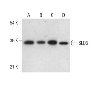
: sc-398784. Western blot analysis of SLD5 expression in A-375 (A), T24 (B), AML-193 (C) and ES-2 (D) whole cell lysates.")
: sc-398784. Fluorescent western blot analysis of SLD5 expression in ES-2 (A) and HeLa (B) whole cell lysates. Blocked with UltraCruz<sup>®</sup> Blocking Reagent: sc-516214. Detection reagent used: m-IgG<sub>1</sub> BP-CFL 647: sc-533664.")
: sc-398784. Western blot analysis of SLD5 expression in ES-2 (A) and HeLa (B) whole cell lysates.")
SLD5 Antibody (D-7): sc-398784
- SLD5 Antibody (D-7) is a mouse monoclonal IgG1 κ SLD5 antibody, cited in 3 publications, provided at 200 µg/ml
- raised against amino acids 1-223 representing full length SLD5 of human origin
- SLD5 Antibody (D-7) is recommended for detection of SLD5 of human origin by WB, IP, IF and ELISA
- Anti-SLD5 Antibody (D-7) is available conjugated to agarose for IP; HRP for WB, IHC(P) and ELISA; and to either phycoerythrin or FITC for IF, IHC(P) and FCM
- also available conjugated to Alexa Fluor® 488, Alexa Fluor® 546, Alexa Fluor® 594 or Alexa Fluor® 647 for WB (RGB), IF, IHC(P) and FCM, and for use with RGB fluorescent imaging systems, such as iBright™ FL1000, FluorChem™, Typhoon, Azure and other comparable systems
- also available conjugated to Alexa Fluor® 680 or Alexa Fluor® 790 for WB (NIR), IF and FCM; for use with Near-Infrared (NIR) detection systems, such as LI-COR®Odyssey®, iBright™ FL1000, FluorChem™, Typhoon, Azure and other comparable systems
- m-IgG Fc BP-HRP and m-IgG1 BP-HRP are the preferred secondary detection reagents for SLD5 Antibody (D-7) for WB applications. These reagents are now offered in bundles with SLD5 Antibody (D-7) (see ordering information below).

QUICK LINKS
SEE ALSO...
SLD5 Antibody (D-7) is a mouse monoclonal IgG1 kappa light chain antibody that detects SLD5 protein of human origin by western blotting (WB), immunoprecipitation (IP), immunofluorescence (IF), and enzyme-linked immunosorbent assay (ELISA). SLD5 (D-7) antibody is available in both non-conjugated and various conjugated forms, including agarose, horseradish peroxidase (HRP), phycoerythrin (PE), fluorescein isothiocyanate (FITC), and multiple Alexa Fluor® conjugates. SLD5 protein, also known as GINS4 (GINS complex subunit 4), is a 223 amino acid protein located in both cytoplasm and nucleus, and plays a pivotal role in DNA replication initiation and progression. As a critical component of GINS complex, which consists of Psf1, Psf2, and Psf3 proteins, SLD5 is essential for proper assembly of this heterotetrameric complex. GINS complex is highly conserved across evolution and crucial for maintaining genomic stability during cell division. Notably, SLD5 has been found significantly up-regulated in aggressive melanomas, highlighting potential role in cancer biology. Interaction between SLD5 and Psf1 is particularly important, as Psf1 binds to single-stranded DNA, facilitating GINS complex′s function in DNA replication. Using anti-SLD5 antibody (D-7), researchers can gain valuable insights into DNA replication mechanisms and SLD5′s implications in various cellular processes and diseases.
Alexa Fluor® is a trademark of Molecular Probes Inc., OR., USA
LI-COR® and Odyssey® are registered trademarks of LI-COR Biosciences
SLD5 Antibody (D-7) References:
- PSF1 is essential for early embryogenesis in mice. | Ueno, M., et al. 2005. Mol Cell Biol. 25: 10528-32. PMID: 16287864
- Identification and characterization of mouse PSF1-binding protein, SLD5. | Kong, L., et al. 2006. Biochem Biophys Res Commun. 339: 1204-7. PMID: 16338220
- Structure of the human GINS complex and its assembly and functional interface in replication initiation. | Kamada, K., et al. 2007. Nat Struct Mol Biol. 14: 388-96. PMID: 17417653
- Molecular architecture of the human GINS complex. | Boskovic, J., et al. 2007. EMBO Rep. 8: 678-84. PMID: 17557111
- Comprehensive expression profiling of tumor cell lines identifies molecular signatures of melanoma progression. | Ryu, B., et al. 2007. PLoS One. 2: e594. PMID: 17611626
- Crystal structure of the GINS complex and functional insights into its role in DNA replication. | Chang, YP., et al. 2007. Proc Natl Acad Sci U S A. 104: 12685-90. PMID: 17652513
- The initiation step of eukaryotic DNA replication. | Pospiech, H., et al. 2010. Subcell Biochem. 50: 79-104. PMID: 20012578
- PSF3 marks malignant colon cancer and has a role in cancer cell proliferation. | Nagahama, Y., et al. 2010. Biochem Biophys Res Commun. 392: 150-4. PMID: 20059967
- Structure and function of the GINS complex, a key component of the eukaryotic replisome. | MacNeill, SA. 2010. Biochem J. 425: 489-500. PMID: 20070258
Ordering Information
| Product Name | Catalog # | UNIT | Price | Qty | FAVORITES | |
SLD5 Antibody (D-7) | sc-398784 | 200 µg/ml | $322.00 | |||
SLD5 Antibody (D-7): m-IgG Fc BP-HRP Bundle | sc-527811 | 200 µg Ab; 10 µg BP | $361.00 | |||
SLD5 Antibody (D-7): m-IgG1 BP-HRP Bundle | sc-533184 | 200 µg Ab; 20 µg BP | $361.00 | |||
SLD5 Antibody (D-7) AC | sc-398784 AC | 500 µg/ml, 25% agarose | $424.00 | |||
SLD5 Antibody (D-7) HRP | sc-398784 HRP | 200 µg/ml | $322.00 | |||
SLD5 Antibody (D-7) FITC | sc-398784 FITC | 200 µg/ml | $336.00 | |||
SLD5 Antibody (D-7) PE | sc-398784 PE | 200 µg/ml | $349.00 | |||
SLD5 Antibody (D-7) Alexa Fluor® 488 | sc-398784 AF488 | 200 µg/ml | $364.00 | |||
SLD5 Antibody (D-7) Alexa Fluor® 546 | sc-398784 AF546 | 200 µg/ml | $364.00 | |||
SLD5 Antibody (D-7) Alexa Fluor® 594 | sc-398784 AF594 | 200 µg/ml | $364.00 | |||
SLD5 Antibody (D-7) Alexa Fluor® 647 | sc-398784 AF647 | 200 µg/ml | $364.00 | |||
SLD5 Antibody (D-7) Alexa Fluor® 680 | sc-398784 AF680 | 200 µg/ml | $364.00 | |||
SLD5 Antibody (D-7) Alexa Fluor® 790 | sc-398784 AF790 | 200 µg/ml | $364.00 |