")
Anticorps EDG-1/S1P1/S1PR1 (A-6): sc-48356
- L'anticorps EDG-1/S1P1/S1PR1 A-6 est un monoclonal IgG3 κ L'Anticorps EDG-1/S1P1/S1PR1, cité dans 23 publications, fourni en 200 µg/ml
- dirigé contre les acides aminés 322-381 de EDG-1 d'origine human
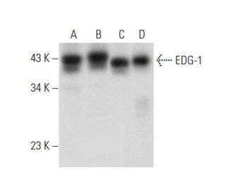
- recommandé pour la détection de EDG-1 d'origine human par WB, IP, IF, IHC(P) et ELISA
- disponible conjugué à l'agarose pour IP; à l'HRP pour WB, IHC(P) et ELISA; à la phycoérythrine, à la fluorescéine, à l'Alexa Fluor® 488 ou à l'Alexa Fluor® 647 pour IF, IHC(P) et FCM
- Nous testons actuellement encore nos anticorps secondaires pour trouver la meilleure protéine de liaison pour cet anticorps primaire EDG-1/S1P1/S1PR1 (A-6). Veuillez nous contacter si vous avez des questions concernant les réactifs de détection secondaire appropriés.

ACCÈS RAPIDE AUX LIENS
VOIR ÉGALEMENT...
L'anticorps EDG-1 (A-6) est un anticorps monoclonal IgG3 à chaîne légère kappa de souris qui détecte l'EDG-1 d'origine humaine par western blotting (WB), immunoprécipitation (IP), immunofluorescence (IF), immunohistochimie et enzyme-linked immunosorbent assay (ELISA). L'anticorps EDG-1 (A-6) est disponible sous forme non conjuguée et sous diverses formes conjuguées, notamment agarose, peroxydase de raifort (HRP), phycoérythrine (PE), isothiocyanate de fluorescéine (FITC) et plusieurs conjugués Alexa Fluor®. EDG-1, également connu sous le nom de S1PR1 ou S1P1, est un membre de la famille du gène de différenciation endothéliale (EDG) des récepteurs couplés aux protéines G, qui jouent un rôle crucial dans la médiation des effets des molécules de signalisation lysophospholipides telles que la sphingosine-1-phosphate (S1P) et l'acide lysophosphatidique (LPA). L'EDG-1 est principalement situé à la surface des cellules et participe à divers processus cellulaires, notamment la survie, la croissance, la migration et la différenciation des cellules, en se couplant aux protéines Gi pour activer les voies de signalisation en aval telles que l'Akt. Ce rôle est particulièrement important dans les cellules de gliome, où l'expression de l'EDG-1 est associée à la progression et à la survie de la tumeur. La capacité de l'EDG-1 à interagir avec de multiples protéines G influence un large éventail de réponses physiologiques, ce qui fait de l'EDG-1 une cible importante pour la recherche en biologie du cancer et le développement thérapeutique.
Alexa Fluor® est une marque déposée de Molecular Probes Inc., OR., USA
LI-COR® et Odyssey® sont marques déposées de LI-COR Biosciences
Anticorps EDG-1/S1P1/S1PR1 (A-6) Références:
- Propriétés pharmacologiques différentielles et transduction du signal des récepteurs de la sphingosine 1-phosphate EDG-1, EDG-3 et EDG-5. | Ancellin, N. and Hla, T. 1999. J Biol Chem. 274: 18997-9002. PMID: 10383399
- Une sous-famille de récepteurs cellulaires couplés aux protéines G pour les lysophospholipides et les lysosphingolipides. | Goetzl, EJ. and An, S. 1999. Adv Exp Med Biol. 469: 259-64. PMID: 10667339
- La sphingosine-1-phosphate est un ligand du récepteur EDG-6 couplé à la protéine G. | Van Brocklyn, JR., et al. 2000. Blood. 95: 2624-9. PMID: 10753843
- Signalisation de la sphingosine-1-phosphate via la famille EDG-1 de récepteurs couplés à la protéine G. | Hla, T., et al. 2000. Ann N Y Acad Sci. 905: 16-24. PMID: 10818438
- Sphingosine 1-phosphate: un ligand pour la famille EDG-1 de récepteurs couplés à la protéine G. | Spiegel, S. 2000. Ann N Y Acad Sci. 905: 54-60. PMID: 10818441
- Signalisation de la sphingosine 1-phosphate dans les cellules de mammifères. | Pyne, S. and Pyne, NJ. 2000. Biochem J. 349: 385-402. PMID: 10880336
- Rôles différentiels de Edg-1 et Edg-5, récepteurs de la sphingosine 1-phosphate, dans les voies de signalisation des cellules de gliome C6. | Sato, K., et al. 2000. Brain Res Mol Brain Res. 85: 151-60. PMID: 11146117
- Effets sélectifs des récepteurs de l'acide lysophosphatidique sur la migration des cellules T Jurkat à travers la membrane basale d'un modèle de Matrigel. | Zheng, Y., et al. 2001. J Immunol. 166: 2317-22. PMID: 11160288
- Distribution des récepteurs Edg2 dans le cerveau du rat adulte. | Handford, EJ., et al. 2001. Neuroreport. 12: 757-60. PMID: 11277579
- La sphingosine 1-phosphate active l'Akt, la production d'oxyde nitrique et la chimiotaxie par le biais d'une voie protéine Gi/phosphoinositide 3-kinase dans les cellules endothéliales. | Morales-Ruiz, M., et al. 2001. J Biol Chem. 276: 19672-7. PMID: 11278592
- Rôles de la N-glycosylation dans la dynamique de Edg-1/S1P1 dans les cellules stimulées par la sphingosine 1-phosphate. | Kohno, T. and Igarashi, Y. 2004. Glycoconj J. 21: 497-501. PMID: 15750791
Informations pour la commande
| Nom du produit | Ref. Catalogue | COND. | Prix HT | QTÉ | Favoris | |
Anticorps EDG-1/S1P1/S1PR1 (A-6) | sc-48356 | 200 µg/ml | $322.00 | |||
Anticorps EDG-1/S1P1/S1PR1 (A-6) AC | sc-48356 AC | 500 µg/ml, 25% agarose | $424.00 | |||
Anticorps EDG-1/S1P1/S1PR1 (A-6) Alexa Fluor® 488 | sc-48356 AF488 | 200 µg/ml | $364.00 | |||
Anticorps EDG-1/S1P1/S1PR1 (A-6) Alexa Fluor® 647 | sc-48356 AF647 | 200 µg/ml | $364.00 | |||
Anticorps EDG-1/S1P1/S1PR1 (A-6) FITC | sc-48356 FITC | 200 µg/ml | $336.00 | |||
Anticorps EDG-1/S1P1/S1PR1 (A-6) HRP | sc-48356 HRP | 200 µg/ml | $322.00 | |||
Anticorps EDG-1/S1P1/S1PR1 (A-6) PE | sc-48356 PE | 200 µg/ml | $349.00 |