Date published: 2025-11-21

1-800-457-3801

SCBT Portrait Logo
Seach Input

Visual Arrestin Antibody (B-2): sc-166068

4.5(4)
Write a reviewAsk a question

Datasheets
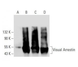
  • Visual Arrestin Antibody (B-2) is a mouse monoclonal IgG3 κ provided at 200 µg/ml
  • raised against amino acids 316-405 mapping at the C-terminus of Visual Arrestin of human origin
  • recommended for detection of Visual Arrestin of mouse, rat and human origin by WB, IP, IF, IHC(P) and ELISA
  • m-IgG3 BP-HRP is the preferred secondary detection reagent for Visual Arrestin Antibody (B-2) for WB and IHC(P) applications. This reagent is now offered in a bundle with Visual Arrestin Antibody (B-2) (see ordering information below).

    QUICK LINKS

    SEE ALSO...

    Visual Arrestin Antibody (B-2) is a mouse monoclonal IgG3 kappa light chain antibody designed to detect Visual Arrestin, also known as Arrestin 1, retinal S-antigen, or S-Arrestin, across multiple species including human, mouse, and rat. anti-Visual Arrestin antibody (B-2) is raised against amino acids 316-405 at the C-terminus of the human Visual Arrestin protein, ensuring effective binding for reliable detection in applications such as western blotting (WB), immunoprecipitation (IP), immunofluorescence (IF), immunohistochemistry with paraffin-embedded sections (IHCP), and enzyme-linked immunosorbent assay (ELISA). Visual Arrestin plays a critical role in the phototransduction pathway by binding to phosphorylated rhodopsin, thereby terminating the light signal and preventing continuous activation of G protein-coupled receptors. This function is essential for maintaining accurate visual perception and preventing signal desensitization, allowing organisms to respond appropriately to changes in light conditions. Additionally, Visual Arrestin is expressed in the pineal gland, where Visual Arrestin contributes to the regulation of circadian rhythms, highlighting Visual Arrestin′s importance in both vision and overall physiological regulation. The ability of Visual Arrestin to modulate receptor activity makes Visual Arrestin a key player in cellular responses to light and other stimuli, influencing processes such as neurotransmission and cellular adaptation. Visual Arrestin (B-2) antibody facilitates detailed studies of Visual Arrestin′s function and regulation, aiding research into visual signal transduction, retinal diseases, and autoimmune conditions like uveoretinitis and Oguchi disease.

    For Research Use Only. Not Intended for Diagnostic or Therapeutic Use.

    Ordering Information

    Product NameCatalog #UNITPriceQtyFAVORITES

    Visual Arrestin Antibody (B-2)

    sc-166068
    200 µg/ml
    $316.00

    Visual Arrestin Antibody (B-2): m-IgG3 BP-HRP Bundle

    sc-550389
    200 µg Ab; 40 µg BP
    $354.00

    Can Visual Arrestin (B-2): sc-166068 monoclonal antibody be used to stain formalin-fixed, paraffin-embedded (FFPE) tissue sections?

    Asked by: cjMara
    Thank you for your question. Yes, Visual Arrestin (B-2): sc-166068 has been validated for use in IHC with paraffin-embedded sections. We recommend performing antigen retrieval with sodium citrate buffer (pH 6) and heat. The full protocol can be found here: https://www.scbt.com/scbt/resources/protocols/immunoperoxidase-staining
    Answered by: Technical Support
    Date published: 2017-02-27
    • y_2025, m_11, d_13, h_10CST
    • bvseo_bulk, prod_bvqa, vn_bulk_3.0.42
    • cp_1, bvpage1
    • co_hasquestionsanswers, tq_1
    • loc_en_US, sid_166068, prod, sort_[SortEntry(order=LAST_APPROVED_ANSWER_SUBMISSION_TIME, direction=DESCENDING)]
    • clientName_scbt
    • bvseo_sdk, java_sdk, bvseo-4.0.0
    • CLOUD, getContent, 117ms
    • QUESTIONS, PRODUCT
    Rated 5 out of 5 by from Good specificity shown by using transfectedGood specificity shown by using transfected and non-transfected 293T cells for over-expression of the human Visual Arrestin protein via Western Blot analysis. -SCBT QC
    Date published: 2015-03-18
    Rated 4 out of 5 by from WB positive with ARPEWB positive with ARPE-19 and RPE-J cells. Some non-specific bands also observed. -SCBT QC
    Date published: 2015-01-12
    Rated 4 out of 5 by from Positive detection of the Visual ArrestinPositive detection of the Visual Arrestin protein by WB using mouse and rat eye tissue extracts. Large band at expected MW observed; numerous non-specific bands observed as well (poss. due to over-exposure or amount of protein/antibody used). -SCBT QC
    Date published: 2015-01-05
    Rated 5 out of 5 by from An increase in signal was observed via WesternAn increase in signal was observed via Western Blot when comparing mouse 293T cells transfected with mouse Visual Arrestin protein with those that have not. -SCBT QC
    Date published: 2013-08-17
    • y_2025, m_11, d_13, h_10
    • bvseo_bulk, prod_bvrr, vn_bulk_3.0.42
    • cp_1, bvpage1
    • co_hasreviews, tv_0, tr_4
    • loc_en_US, sid_166068, prod, sort_[SortEntry(order=SUBMISSION_TIME, direction=DESCENDING)]
    • clientName_scbt
    • bvseo_sdk, java_sdk, bvseo-4.0.0
    • CLOUD, getReviews, 15ms
    • REVIEWS, PRODUCT
    Visual Arrestin Antibody (B-2) is rated 4.5 out of 5 by 4.
    • y_2025, m_11, d_13, h_10
    • bvseo_bulk, prod_bvrr, vn_bulk_3.0.42
    • cp_1, bvpage1
    • co_hasreviews, tv_0, tr_4
    • loc_en_US, sid_166068, prod, sort_[SortEntry(order=SUBMISSION_TIME, direction=DESCENDING)]
    • clientName_scbt
    • bvseo_sdk, java_sdk, bvseo-4.0.0
    • CLOUD, getAggregateRating, 111ms
    • REVIEWS, PRODUCT