")
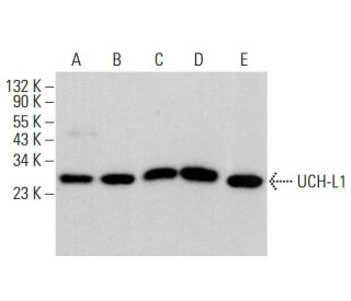
UCH-L1 Antikörper (13C4): sc-58594
- UCH-L1 Antikörper (13C4) ist ein Maus monoklonales IgG2a κ, verwendet in 3 wissenschaftlichen Veröffentlichungen, in einer Menge von 200 µg/ml
- gegen natives UCH-L1 Protein human Herkunft
- Empfohlen für die Detektion von UCH-L1 on neuronal cell bodies and axons in the CNS and periphery, small nerve fibers in peripheral tissues, neuroendocrine cells in the pituitary, thyroid, pancreas and tumors of the DNES aus der Spezies mouse, rat und human per WB, IP, IF und IHC(P); auch empfohlen für den Nachweis von UCH-L1 in human adult gut
- m-IgGκ BP-HRP ist das bevorzugte sekundäre Nachweisreagenz für UCH-L1 Antikörper (13C4) für WB- und IHC(P)-Anwendungen. Dieses Reagenz wird jetzt in einem Paket mit UCH-L1 Antikörper (13C4) angeboten(siehe Bestellinformationen unten). Weitere m-IgGκ BP-Konjugate finden Sie in unserer vollständigen Liste der Maus-IgG-Bindungsproteine.

UCH-L1 (Ubiquitin C-terminal Hydrolase) ist ein Mitglied einer Genfamilie, deren Produkte kleine C-terminale Addukte von Ubiquitin hydrolysieren, um das Ubiquitin Monomer zu generieren. Die Expression von UCH-L1 ist sehr spezifisch für Neuronen und Zellen des diffusen neuroendokrinen Systems und deren Tumoren. UCH-L1 wird in Gehirnneuronen exprimiert. Eine Untersuchung bestimmter Gehirnregionen zeigt eine Expression in allen getesteten Bereichen, insbesondere in der Substantia nigra. UCH-L1 stellt 1-2% der totalen löslichen Gehirnproteine dar. Sein Auftreten in Lewy-Körpern und seine Funktion in der Proteasom-Pathway machen es zu einem überzeugenden Kandidatengen in der Parkinson-Krankheit. Das Gen, das UCH-L1 codiert, wird auf dem menschlichen Chromosom 4p13 abgebildet. Das 230 Aminosäure lange menschliche UCH-L3-Protein ist 54% identisch mit dem von UCH-L1. UCH-L3 ist die vorherrschende Thiol-Protease und hat hochaffine Bindungsstellen für Ubiquitin.
Alexa Fluor® ist ein Markenzeichen von Molecular Probes Inc., OR., USA
LI-COR® und Odyssey® sind Markenzeichen von LI-COR Biosciences
UCH-L1 Antikörper (13C4) Literaturhinweise:
- Die Funktionen von UCH-L1 und seine Beziehung zu neurodegenerativen Erkrankungen. | Setsuie, R. and Wada, K. 2007. Neurochem Int. 51: 105-11. PMID: 17586089
- Familiäre Mutationen und posttranslationale Modifikationen von UCH-L1 bei der Parkinson-Krankheit und neurodegenerativen Erkrankungen. | Lee, YC. and Hsu, SD. 2017. Curr Protein Pept Sci. 18: 733-745. PMID: 26899237
- Zeitlicher Verlauf und diagnostische Genauigkeit der glialen und neuronalen Blut-Biomarker GFAP und UCH-L1 in einer großen Kohorte von Traumapatienten mit und ohne mildem Schädel-Hirn-Trauma. | Papa, L., et al. 2016. JAMA Neurol. 73: 551-60. PMID: 27018834
- Ubiquitin C-terminale Hydrolase-L1 (UCH-L1) als therapeutisches und diagnostisches Ziel bei Neurodegeneration, Neurotrauma und Neuroverletzungen. | Wang, KK., et al. 2017. Expert Opin Ther Targets. 21: 627-638. PMID: 28434268
- Ubiquitin C-Terminal Hydrolase-L1 (UCH-L1) in der Vorhersage von Computertomographie-Befunden bei traumatischen Hirnverletzungen; eine Meta-Analyse. | Ramezani, F., et al. 2018. Emerg (Tehran). 6: e62. PMID: 30788389
- S100B, GFAP, UCH-L1 und NSE als Prädiktoren für Anomalien bei der CT-Bildgebung nach leichten traumatischen Hirnverletzungen: eine systematische Überprüfung und Metaanalyse der diagnostischen Testgenauigkeit. | Amoo, M., et al. 2022. Neurosurg Rev. 45: 1171-1193. PMID: 34709508
- UCH-L1 und UCH-L3 regulieren die krebsstammzellähnlichen Eigenschaften durch den PI3 K/Akt-Signalweg in Prostatakrebszellen. | Lee, JE., et al. 2021. Anim Cells Syst (Seoul). 25: 312-322. PMID: 34745437
- Prognostischer Wert der GFAP- und UCH-L1-Konzentrationen am Tag der Verletzung für die Vorhersage der funktionellen Erholung nach einem Schädel-Hirn-Trauma bei Patienten aus der US-amerikanischen TRACK-TBI-Kohorte: eine beobachtende Kohortenstudie. | Korley, FK., et al. 2022. Lancet Neurol. 21: 803-813. PMID: 35963263
- Dysfunktionales UCH-L1 hemmt die Proteostase. | Wang, M. 2023. Nat Rev Nephrol. 19: 424. PMID: 37258710
- Die Proximity-Proteomik zeigt, dass UCH-L1 ein wesentlicher Regulator der NLRP3-vermittelten IL-1β-Produktion in menschlichen Makrophagen und Mikroglia ist. | Liang, Z., et al. 2024. Cell Rep. 43: 114152. PMID: 38669140
Bestellinformation
| Produkt | Katalog # | EINHEIT | Preis | ANZAHL | Favoriten | |
UCH-L1 Antikörper (13C4) | sc-58594 | 200 µg/ml | $322.00 | |||
UCH-L1 (13C4): m-IgGκ BP-HRP Bundle | sc-521055 | 200 µg Ab, 40 µg BP | $361.00 |