")
TAP1 Antibody (B-8): sc-376796
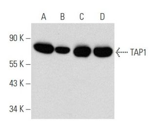
- TAP1 Antibody (B-8) is a mouse monoclonal IgG3 κ TAP1 antibody, cited in 3 publications, provided at 200 µg/ml
- specific for an epitope mapping between amino acids 697-724 at the C-terminus of TAP1 of mouse origin
- recommended for detection of all TAP1 isoforms of mouse, rat and human origin by WB, IP, IF, IHC(P) and ELISA
- m-IgGκ BP-HRP is the preferred secondary detection reagent for TAP1 Antibody (B-8) for WB and IHC(P) applications. This reagent is now offered in a bundle with TAP1 Antibody (B-8) (see ordering information below). For additional m-IgGκ BP conjugates see our complete list of Mouse IgG Binding Proteins.

QUICK LINKS
TAP1 Antibody (B-8) is a mouse monoclonal IgG3 kappa light chain antibody that detects TAP1 protein of mouse, rat, and human origin by western blotting (WB), immunoprecipitation (IP), immunofluorescence (IF), immunohistochemistry, and enzyme-linked immunosorbent assay (ELISA). TAP1 (B-8) antibody is available as the non-conjugated form. The transporter associated with antigen processing (TAP) plays a crucial role in the immune response by facilitating peptide transport from the cytosol into the endoplasmic reticulum (ER) for loading onto major histocompatibility complex (MHC) class I molecules, which are essential for presenting antigens to CD8+ T cells. This process is vital for recognizing and eliminating infected or malignant cells, making TAP1 a key player in adaptive immunity. The TAP transporter consists of two subunits, TAP1 and TAP2, forming a dimeric structure that creates a translocation pore within the ER membrane. Each subunit contains a transmembrane region contributing to pore formation, as well as a cytoplasmic peptide-binding pocket ensuring selective peptide transport. Additionally, TAP1 and TAP2 possess nucleotide-binding domains that hydrolyze ATP, providing energy for conformational changes that facilitate peptide translocation. Proper functioning of TAP1 remains critical for effective immune surveillance and response, highlighting its significance in both basic immunology and potential therapeutic applications.
Alexa Fluor® is a trademark of Molecular Probes Inc., OR., USA
LI-COR® and Odyssey® are registered trademarks of LI-COR Biosciences
TAP1 Antibody (B-8) References:
- Nucleotide binding by TAP mediates association with peptide and release of assembled MHC class I molecules. | Knittler, MR., et al. 1999. Curr Biol. 9: 999-1008. PMID: 10508608
- Membrane topology and dimerization of the two subunits of the transporter associated with antigen processing reveal a three-domain structure. | Vos, JC., et al. 1999. J Immunol. 163: 6679-85. PMID: 10586064
- Pairing of the nucleotide binding domains of the transporter associated with antigen processing. | Lapinski, PE., et al. 2000. J Biol Chem. 275: 6831-40. PMID: 10702242
- Characteristics of peptide and major histocompatibility complex class I/beta 2-microglobulin binding to the transporters associated with antigen processing (TAP1 and TAP2). | Androlewicz, MJ., et al. 1994. Proc Natl Acad Sci U S A. 91: 12716-20. PMID: 7809108
- Evidence that transporters associated with antigen processing translocate a major histocompatibility complex class I-binding peptide into the endoplasmic reticulum in an ATP-dependent manner. | Androlewicz, MJ., et al. 1993. Proc Natl Acad Sci U S A. 90: 9130-4. PMID: 8415666
- Multiple regions of the transporter associated with antigen processing (TAP) contribute to its peptide binding site. | Nijenhuis, M. and Hämmerling, GJ. 1996. J Immunol. 157: 5467-77. PMID: 8955196
- Major histocompatibility complex class I molecules interact with both subunits of the transporter associated with antigen processing, TAP1 and TAP2. | Powis, SJ. 1997. Eur J Immunol. 27: 2744-7. PMID: 9368636
Ordering Information
| Product Name | Catalog # | UNIT | Price | Qty | FAVORITES | |
TAP1 Antibody (B-8) | sc-376796 | 200 µg/ml | $322.00 | |||
TAP1 Antibody (B-8): m-IgGκ BP-HRP Bundle | sc-523096 | 200 µg Ab, 40 µg BP | $361.00 | |||
TAP1 (B-8) Neutralizing Peptide | sc-376796 P | 100 µg/0.5 ml | $69.00 |