
RIP3 Lentiviral Activation Particles (m): sc-425224-LAC
- Target species: mouse
- 200 µl of transduction-ready, high-titer CRISPR/dCas9 Lentiviral Activation Particles
- RIP3 Lentiviral Activation Particles (m) is a synergistic activation mediator (SAM) transcription activation system designed to specifically and efficiently upregulate gene expression via lentiviral transduction of cells
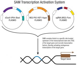
- RIP3 Lentiviral Activation Particles (m) contain the following SAM Activation elements: a deactivated Cas9 (dCas9) nuclease (D10A and N863A) fused to the transactivation domain VP64, an MS2-p65-HSF1 fusion protein and a target-specific 20 nt guide RNA. They also contain the blasticidin, hygromycin and puromycin resistance genes
- Upon transduction, the SAM complex binds to a site-specific region approximately 200-250 nt upstream of the transcriptional start site and provides robust recruitment of transcription factors for highly efficient gene activation
- gRNAs encoded by RIP3 Lentiviral Activation Plasmid (m) and RIP3 Lentiviral Activation Plasmid (m2) target distinct regulatory regions of the Ripk3 promoter. One or both designs may be available
- Following transfection, gene activation efficiency can be assayed by WB, IF or IHC using antibody: RIP3 Antibody (B-2): sc-374639

Ordering Information
| Product Name | Catalog # | UNIT | Price | Qty | FAVORITES | |
RIP3 Lentiviral Activation Particles (m) | sc-425224-LAC | 200 µl | $455.00 | |||
RIP3 Lentiviral Activation Particles (m2) | sc-425224-LAC-2 | 200 µl | $455.00 |
Mouse Ripk3 encodes the serine/threonine kinase RIP3, a central regulator of programmed necrosis (necroptosis) downstream of death receptors and innate immune sensing. RIP3 functions in the RIPK1–RIPK3 signaling axis to promote MLKL phosphorylation and membrane disruption, integrating inputs from TNF, TLRs, and interferon-stimulated pathways. Through cross-talk with apoptosis and inflammatory signaling, RIP3 influences cytokine production, tissue injury responses, and host–pathogen interactions. Dysregulated RIP3 activity has been implicated in models of inflammatory and neurodegenerative pathology, ischemic damage, and infection-associated tissue injury, making it a key node for mechanistic studies of cell death and inflammation.
RIP3 Lentiviral Activation Particles (m) address this need by packaging the complete synergistic activation mediator (SAM) transcriptional activation system into transduction-ready, high-titer lentiviral particles, enabling efficient Ripk3 upregulation across a broader range of human cell types.
RIP3 Lentiviral Activation Particles (m) deliver all functional components of the synergistic activation mediator (SAM) system via lentiviral transduction. The system comprises three particle preparations co-transduced into target cells: one encoding catalytically inactive dCas9 (D10A and N863A mutations) fused to the VP64 transactivation domain with a blasticidin resistance gene; one encoding the MS2-p65-HSF1 fusion protein with a hygromycin resistance gene; and one encoding a target-specific 20 nt sgRNA fused to two MS2 RNA aptamers with a puromycin resistance gene. Following lentiviral transduction and genomic integration of the expression cassettes, the SAM components are stably expressed and assemble at the target locus within the proximal promoter region upstream of the Ripk3 transcriptional start site, where VP64, p65, and HSF1 act cooperatively to recruit endogenous transcriptional machinery and drive sustained upregulation of endogenous RIP3 expression. The use of nuclease-inactive dCas9 avoids the introduction of double-strand DNA breaks and preserves the native Ripk3 genomic locus and regulatory architecture.
The lentiviral format offers several practical advantages: stable genomic integration supports heritable activation across cell divisions; high-titer particle preparations eliminate the need for in-house viral production; and compatibility with primary, non-dividing, and transfection-resistant cell types expands experimental accessibility. Successful transduction can be confirmed and enriched through triple antibiotic selection using puromycin, hygromycin, and blasticidin.
For Research Use Only. Not Intended for Diagnostic or Therapeutic Use.