Date published: 2026-1-11

1-800-457-3801

SCBT Portrait Logo
Seach Input

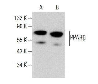
PPARβ Antibody (F-7): sc-74440

4.4(7)
Write a reviewAsk a question

Datasheets
  • PPARβ Antibody (F-7) is a mouse monoclonal IgG1 κ, cited in 9 publications, provided at 200 µg/ml
  • raised against amino acids 2-75 of PPARβ of human origin
  • recommended for detection of PPARβ of human origin by WB, IP, IF, IHC(P) and ELISA
  • See PPARβ (F-10): sc-74517 for PPARβ antibody conjugates, including AC, HRP, FITC, PE, Alexa Fluor® 488, 594, 647, 680 and 790.
  • m-IgG Fc BP-HRP and m-IgG1 BP-HRP are the preferred secondary detection reagents for PPARβ Antibody (F-7) for WB and IHC(P) applications. These reagents are now offered in bundles with PPARβ Antibody (F-7) (see ordering information below).

QUICK LINKS

SEE ALSO...

PPARβ Antibody (F-7) is a mouse monoclonal IgG1 antibody that detects PPARβ in human samples through various applications including western blotting (WB), immunoprecipitation (IP), immunofluorescence (IF), immunohistochemistry with paraffin-embedded sections (IHCP), and enzyme-linked immunosorbent assay (ELISA). PPARβ, also known as PPARδ, is a crucial member of the peroxisome proliferator-activated receptor family, which plays a significant role in regulating gene expression related to lipid metabolism, energy homeostasis, and inflammation. Anti-PPARβ antibody (F-7) targets a protein predominantly located in the nucleus, where PPARβ functions as a transcription factor by forming heterodimers with retinoid X receptors and binding to specific DNA sequences known as PPAR-response elements. Nuclear localization of PPARβ is vital for mediating the effects of fatty acids and other ligands on gene transcription, influencing metabolic processes and cellular differentiation. PPARβ is widely expressed across various tissues, making PPARβ an important target for studying metabolic disorders and potential therapeutic interventions. PPARβ monoclonal antibody (F-7) serves as an essential tool for researchers investigating the intricate pathways regulated by PPARβ and its implications in health and disease.

For Research Use Only. Not Intended for Diagnostic or Therapeutic Use.

Ordering Information

Product NameCatalog #UNITPriceQtyFAVORITES

PPARβ Antibody (F-7)

sc-74440
200 µg/ml
$322.00

PPARβ Antibody (F-7): m-IgG Fc BP-HRP Bundle

sc-539519
200 µg Ab; 10 µg BP
$361.00

PPARβ Antibody (F-7): m-IgG1 BP-HRP Bundle

sc-541540
200 µg Ab; 20 µg BP
$361.00

I am using PPARβ (F-7): sc-74440 monoclonal antibody to stain paraffin embedded tissue. Do you recommend antigen retrieval when using this antibody?

Asked by: Professor Griffin
We recommend performing antigen retrieval with sodium citrate buffer (pH 6) and heat. The full protocol can be found here: https://www.scbt.com/scbt/resources/protocols/immunoperoxidase-staining
Answered by: Technical Support
Date published: 2017-02-24
  • y_2025, m_12, d_30, h_8CST
  • bvseo_bulk, prod_bvqa, vn_bulk_3.0.42
  • cp_1, bvpage1
  • co_hasquestionsanswers, tq_1
  • loc_en_US, sid_74440, prod, sort_[SortEntry(order=LAST_APPROVED_ANSWER_SUBMISSION_TIME, direction=DESCENDING)]
  • clientName_scbt
  • bvseo_sdk, java_sdk, bvseo-4.0.0
  • CLOUD, getContent, 111ms
  • QUESTIONS, PRODUCT
Rated 5 out of 5 by from CleanClean, crisp bands observed by Western Blot, showing an increase in human-derived cells (HCECs) PPAR expression following treatment with cytokines. -SCBT Publication Review
Date published: 2015-04-30
Rated 4 out of 5 by from By immunohistochemistry (formalin fixedBy immunohistochemistry (formalin fixed, paraffin embedded), nuclear staining was observed in trophoblastic cells of human placenta tissue. -SCBT QC
Date published: 2015-04-02
Rated 4 out of 5 by from Quiescent HPASMCs were treated with PDGFQuiescent HPASMCs were treated with PDGF for various amounts of time, up to 72 hours, then extracted for analysis by Western Blot. Using the sc-74440 antibody, an increase in PPAR expression was observed. -SCBT Publication Review
Date published: 2015-04-01
Rated 5 out of 5 by from Two strong bands at the correct molecularTwo strong bands at the correct molecular weights (Isoform 1 & 2) of PPAR observed by Western Blot for Jurkat whole cell lysate. -SCBT QC
Date published: 2015-03-27
Rated 5 out of 5 by from Beautiful staining by immunofluorescenceBeautiful staining by immunofluorescence demonstrating the expression of PPAR in rat corneal epithelia during wound healing. Good staining as well on samples after treatment with GW501516, a PPAR agonist. -SCBT Publication Review
Date published: 2015-03-20
Rated 3 out of 5 by from Analysis by Western blotAnalysis by Western blot, it was found that HPASMCs have higher expression levels of PPAR than HPAECs. Bands for both cell lines were observed to be faint. -SCBT Publication Review
Date published: 2015-03-10
Rated 5 out of 5 by from CrispCrisp, clean band observed by Western Blot, using human recombinant PPAR fusion protein. -SCBT QC
Date published: 2014-07-18
  • y_2025, m_12, d_30, h_8
  • bvseo_bulk, prod_bvrr, vn_bulk_3.0.42
  • cp_1, bvpage1
  • co_hasreviews, tv_0, tr_7
  • loc_en_US, sid_74440, prod, sort_[SortEntry(order=SUBMISSION_TIME, direction=DESCENDING)]
  • clientName_scbt
  • bvseo_sdk, java_sdk, bvseo-4.0.0
  • CLOUD, getReviews, 15ms
  • REVIEWS, PRODUCT
PPARβ Antibody (F-7) is rated 4.4 out of 5 by 7.
  • y_2025, m_12, d_30, h_8
  • bvseo_bulk, prod_bvrr, vn_bulk_3.0.42
  • cp_1, bvpage1
  • co_hasreviews, tv_0, tr_7
  • loc_en_US, sid_74440, prod, sort_[SortEntry(order=SUBMISSION_TIME, direction=DESCENDING)]
  • clientName_scbt
  • bvseo_sdk, java_sdk, bvseo-4.0.0
  • CLOUD, getAggregateRating, 101ms
  • REVIEWS, PRODUCT