")
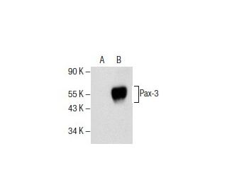
: sc-376204. Western blot analysis of Pax-3 expression in non-transfected: sc-117752 (A) and mouse Pax-3 transfected: sc-122398 (B) 293T whole cell lysates.")
Pax-3 Antibody (F-2): sc-376204
- Pax-3 Antibody (F-2) is a mouse monoclonal IgG3 Pax-3 antibody provided at 200 µg/ml
- specific for an epitope mapping between amino acids 387-425 near the C-terminus of Pax-3 of human origin
- recommended for detection of Pax-3 of mouse, rat, human and origin by WB, IP, IF and ELISA; also reactive with additional species, including and equine, bovine, porcine and avian
- TransCruz reagent for ChIP application (sc-376204 X, 200 µg/0.1 ml)
- See Pax-3/7 (B-5): sc-365843 for Pax-3 antibody conjugates, including AC, HRP, FITC, PE, Alexa Fluor® 488, 594, 647, 680 and 790.
- At present, we have not yet completed the identification of the preferred secondary detection reagent(s) for Pax-3 Antibody (F-2). This work is in progress.

Pax-3 Antibody (F-2) is a mouse monoclonal IgG3 antibody that detects Pax-3 protein of mouse, rat, and human origin by western blotting (WB), immunoprecipitation (IP), immunofluorescence (IF), and enzyme-linked immunosorbent assay (ELISA). Anti-Pax-3 antibody (F-2) is available as the non-conjugated form, which is crucial for studying Pax-3′s role in developmental biology. Pax-3 protein is a vital transcription factor that plays a significant role in early neurogenesis, particularly in the development of the central nervous system. Pax-3 contains both a paired domain and a paired-type homeodomain, which are essential for DNA-binding capabilities, allowing regulation of target genes involved in cell differentiation and proliferation. Pax-3 expression is primarily localized to mitotic cells in the ventricular zone of the developing spinal cord, as well as in specific regions of the hindbrain, midbrain, and diencephalon. During embryonic development, Pax-3 is also expressed in neural crest cells, which are critical for the formation of various structures, including the craniofacial mesectoderm and limb mesenchyme. Mutations in Pax-3 have been linked to Waardenburg syndrome, a genetic condition characterized by hearing loss and pigmentation abnormalities, highlighting Pax-3′s importance in both normal development and disease.
Alexa Fluor® is a trademark of Molecular Probes Inc., OR., USA
LI-COR® and Odyssey® are registered trademarks of LI-COR Biosciences
Pax-3 Antibody (F-2) References:
- Waardenburg's syndrome patients have mutations in the human homologue of the Pax-3 paired box gene. | Tassabehji, M., et al. 1992. Nature. 355: 635-6. PMID: 1347148
- Gene expression signatures identify rhabdomyosarcoma subtypes and detect a novel t(2;2)(q35;p23) translocation fusing PAX3 to NCOA1. | Wachtel, M., et al. 2004. Cancer Res. 64: 5539-45. PMID: 15313887
- Pax-3, a novel murine DNA binding protein expressed during early neurogenesis. | Goulding, MD., et al. 1991. EMBO J. 10: 1135-47. PMID: 2022185
- Autophagy-dependent crosstalk between GILT and PAX-3 influences radiation sensitivity of human melanoma cells. | Hathaway-Schrader, JD., et al. 2018. J Cell Biochem. 119: 2212-2221. PMID: 28857256
- The regenerative potential of Pax3/Pax7 on skeletal muscle injury. | Azhar, M., et al. 2022. J Genet Eng Biotechnol. 20: 143. PMID: 36251225
- Loss of Pax3 causes reduction of melanocytes in the developing mouse cochlea. | Udagawa, T., et al. 2024. Sci Rep. 14: 2210. PMID: 38278860
- Isolation of two isoforms of the PAX3 gene transcripts and their tissue-specific alternative expression in human adult tissues. | Tsukamoto, K., et al. 1994. Hum Genet. 93: 270-4. PMID: 7545913
- Regulation of Pax-3 expression in the dermomyotome and its role in muscle development. | Goulding, M., et al. 1994. Development. 120: 957-71. PMID: 7600971
- Chromosomal localization of seven PAX genes and cloning of a novel family member, PAX-9. | Stapleton, P., et al. 1993. Nat Genet. 3: 292-8. PMID: 7981748
- Mutations in the paired domain of the human PAX3 gene cause Klein-Waardenburg syndrome (WS-III) as well as Waardenburg syndrome type I (WS-I). | Hoth, CF., et al. 1993. Am J Hum Genet. 52: 455-62. PMID: 8447316
- Epistatic relationship between Waardenburg syndrome genes MITF and PAX3. | Watanabe, A., et al. 1998. Nat Genet. 18: 283-6. PMID: 9500554
- The role of Pax3 and Pax7 in development and cancer. | Mansouri, A. 1998. Crit Rev Oncog. 9: 141-9. PMID: 9973247
Ordering Information
| Product Name | Catalog # | UNIT | Price | Qty | FAVORITES | |
Pax-3 Antibody (F-2) | sc-376204 | 200 µg/ml | $322.00 | |||
Pax-3 (F-2) Neutralizing Peptide | sc-376204 P | 100 µg/0.5 ml | $69.00 | |||
Pax-3 Antibody (F-2) X | sc-376204 X | 200 µg/0.1 ml | $322.00 |