")
: sc-514393. Western blot analysis of Paraplegin expression in Hep G2 (A), MIA PaCa-2 (B) and A549 (C) whole cell lysates.")
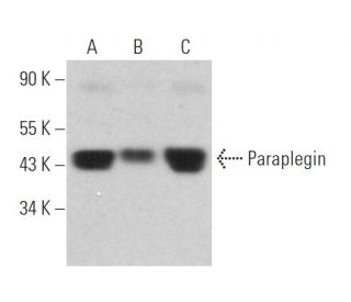
: sc-514393. Western blot analysis of Paraplegin expression in HeLa (A), Hep G2 (B) and JAR (C) whole cell lysates.")
: sc-514393. Western blot analysis of Paraplegin expression in BC3H1 (A), Neuro-2A (B) and PC-12 (C) whole cell lysates.")
Paraplegin Antibody (C-5): sc-514393
- Paraplegin Antibody (C-5) is a mouse monoclonal IgG2b κ Paraplegin antibody, cited in 5 publications, provided at 200 µg/ml
- raised against amino acids 131-310 mapping within an internal region of Paraplegin of human origin
- Paraplegin Antibody (C-5) is recommended for detection of Paraplegin of mouse, rat and human origin by WB, IP, IF and ELISA
- Anti-Paraplegin Antibody (C-5) is available conjugated to agarose for IP; HRP for WB, IHC(P) and ELISA; and to either phycoerythrin or FITC for IF, IHC(P) and FCM
- also available conjugated to Alexa Fluor® 488, Alexa Fluor® 546, Alexa Fluor® 594 or Alexa Fluor® 647 for WB (RGB), IF, IHC(P) and FCM, and for use with RGB fluorescent imaging systems, such as iBright™ FL1000, FluorChem™, Typhoon, Azure and other comparable systems
- also available conjugated to Alexa Fluor® 680 or Alexa Fluor® 790 for WB (NIR), IF and FCM; for use with Near-Infrared (NIR) detection systems, such as LI-COR®Odyssey®, iBright™ FL1000, FluorChem™, Typhoon, Azure and other comparable systems
- At present, we have not yet completed the identification of the preferred secondary detection reagent(s) for Paraplegin Antibody (C-5). This work is in progress.

QUICK LINKS
SEE ALSO...
Paraplegin Antibody (C-5) is a mouse monoclonal IgG2b kappa light chain antibody that detects Paraplegin protein of mouse, rat, and human origin by western blotting (WB), immunoprecipitation (IP), immunofluorescence (IF), and enzyme-linked immunosorbent assay (ELISA). Anti-Paraplegin antibody (C-5) is available in both non-conjugated and various conjugated forms, including agarose, horseradish peroxidase (HRP), phycoerythrin (PE), fluorescein isothiocyanate (FITC), and multiple Alexa Fluor® conjugates. Paraplegin, also known as SPG7 (spastic paraplegia protein 7), CAR, CMAR, or PGN, is a 795 amino acid metalloprotease belonging to the AAA protein family. Paraplegin is primarily localized to the mitochondrial membrane, where Paraplegin plays a crucial role in mitochondrial function and integrity. Paraplegin′s location in the cell is vital for essential processes such as signal transduction and chaperone-like activities within the mitochondria. Defects in the gene encoding Paraplegin are linked to spastic paraplegia type 7 (SPG7), a form of autosomal recessive hereditary spastic paraplegia characterized by muscle spasms, stiffness in the legs, and, in some cases, incontinence. Recent research indicates that SPG7 may represent a mitochondrial-based disease, with mutations in the Paraplegin gene leading to mitochondrial dysfunctions such as ragged-red fibers and impaired axonal transport, ultimately resulting in neurodegeneration associated with hereditary spastic paraplegias.
Alexa Fluor® is a trademark of Molecular Probes Inc., OR., USA
LI-COR® and Odyssey® are registered trademarks of LI-COR Biosciences
Paraplegin Antibody (C-5) References:
- Genomic structure and expression analysis of the spastic paraplegia gene, SPG7. | Settasatian, C., et al. 1999. Hum Genet. 105: 139-44. PMID: 10480368
- A clinical, genetic and biochemical study of SPG7 mutations in hereditary spastic paraplegia. | Wilkinson, PA., et al. 2004. Brain. 127: 973-80. PMID: 14985266
- Mitochondrial proteins in neuronal degeneration. | Lindholm, D., et al. 2004. Biochem Biophys Res Commun. 321: 753-8. PMID: 15358091
- Hereditary spastic paraplegia: respiratory choke or unactivated substrate? | Claypool, SM. and Koehler, CM. 2005. Cell. 123: 183-5. PMID: 16239134
- The m-AAA protease defective in hereditary spastic paraplegia controls ribosome assembly in mitochondria. | Nolden, M., et al. 2005. Cell. 123: 277-89. PMID: 16239145
- Intramuscular viral delivery of paraplegin rescues peripheral axonopathy in a model of hereditary spastic paraplegia. | Pirozzi, M., et al. 2006. J Clin Invest. 116: 202-8. PMID: 16357941
- Mutation analysis of the paraplegin gene (SPG7) in patients with hereditary spastic paraplegia. | Elleuch, N., et al. 2006. Neurology. 66: 654-9. PMID: 16534102
- A novel form of autosomal recessive hereditary spastic paraplegia caused by a new SPG7 mutation. | Warnecke, T., et al. 2007. Neurology. 69: 368-75. PMID: 17646629
- A clinical, genetic, and biochemical characterization of SPG7 mutations in a large cohort of patients with hereditary spastic paraplegia. | Arnoldi, A., et al. 2008. Hum Mutat. 29: 522-31. PMID: 18200586
Ordering Information
| Product Name | Catalog # | UNIT | Price | Qty | FAVORITES | |
Paraplegin Antibody (C-5) | sc-514393 | 200 µg/ml | $322.00 | |||
Paraplegin Antibody (C-5) AC | sc-514393 AC | 500 µg/ml, 25% agarose | $424.00 | |||
Paraplegin Antibody (C-5) HRP | sc-514393 HRP | 200 µg/ml | $322.00 | |||
Paraplegin Antibody (C-5) FITC | sc-514393 FITC | 200 µg/ml | $336.00 | |||
Paraplegin Antibody (C-5) PE | sc-514393 PE | 200 µg/ml | $349.00 | |||
Paraplegin Antibody (C-5) Alexa Fluor® 488 | sc-514393 AF488 | 200 µg/ml | $364.00 | |||
Paraplegin Antibody (C-5) Alexa Fluor® 546 | sc-514393 AF546 | 200 µg/ml | $364.00 | |||
Paraplegin Antibody (C-5) Alexa Fluor® 594 | sc-514393 AF594 | 200 µg/ml | $364.00 | |||
Paraplegin Antibody (C-5) Alexa Fluor® 647 | sc-514393 AF647 | 200 µg/ml | $364.00 | |||
Paraplegin Antibody (C-5) Alexa Fluor® 680 | sc-514393 AF680 | 200 µg/ml | $364.00 | |||
Paraplegin Antibody (C-5) Alexa Fluor® 790 | sc-514393 AF790 | 200 µg/ml | $364.00 |