")
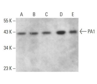
: sc-393387. Western blot analysis of PA1 expression in MCF7 (A), NTERA-2 cl.D1 (B), M1 (C) and F9 (D) whole cell lysates and KNRK nuclear extract (E).")
: sc-393387. Western blot analysis of PA1 expression in non-transfected: sc-117752 (A) and human PA1 transfected: sc-370549 (B) 293T whole cell lysates.")
PA1 Antibody (H-1): sc-393387
- PA1 Antibody (H-1) is a mouse monoclonal IgG1 κ PA1 antibody provided at 200 µg/ml
- raised against amino acids 1-254 representing full length PA1 of human origin
- PA1 Antibody (H-1) is recommended for detection of PA1 of mouse, rat and human origin by WB, IP, IF and ELISA
- Anti-PA1 Antibody (H-1) is available conjugated to agarose for IP; HRP for WB, IHC(P) and ELISA; and to either phycoerythrin or FITC for IF, IHC(P) and FCM
- also available conjugated to Alexa Fluor® 488, Alexa Fluor® 546, Alexa Fluor® 594 or Alexa Fluor® 647 for WB (RGB), IF, IHC(P) and FCM, and for use with RGB fluorescent imaging systems, such as iBright™ FL1000, FluorChem™, Typhoon, Azure and other comparable systems
- also available conjugated to Alexa Fluor® 680 or Alexa Fluor® 790 for WB (NIR), IF and FCM; for use with Near-Infrared (NIR) detection systems, such as LI-COR®Odyssey®, iBright™ FL1000, FluorChem™, Typhoon, Azure and other comparable systems
- m-IgG Fc BP-HRP, m-IgG1 BP-HRP and m-IgGκ BP-HRP are the preferred secondary detection reagents for PA1 Antibody (H-1) for WB applications. These reagents are now offered in bundles with PA1 Antibody (H-1) (see ordering information below).

QUICK LINKS
SEE ALSO...
PA1 Antibody (H-1) is a mouse monoclonal IgG1 kappa light chain antibody that detects PA1 in mouse, rat, and human samples by western blotting (WB), immunoprecipitation (IP), immunofluorescence (IF), and enzyme-linked immunosorbent assay (ELISA). PA1 (H-1) antibody comes in non-conjugated forms and various conjugates, including agarose, horseradish peroxidase (HRP), phycoerythrin (PE), fluorescein isothiocyanate (FITC), and multiple Alexa Fluor® options, offering flexibility for diverse experimental applications. PA1 plays a key role in cellular signaling pathways, especially in regulating cell growth and differentiation, making PA1 essential for understanding various physiological and pathological processes. PA1 function is crucial, as dysregulation can lead to diseases like cancer and neurodegenerative disorders. PA1 primarily resides in the cytoplasm and nucleus, where PA1 interacts with other signaling molecules to influence gene expression and cellular responses to external stimuli. Understanding PA1 localization and function helps reveal disease mechanisms and potential therapeutic targets.
Alexa Fluor® is a trademark of Molecular Probes Inc., OR., USA
LI-COR® and Odyssey® are registered trademarks of LI-COR Biosciences
PA1 Antibody (H-1) References:
- Homozygosity mapping of giant axonal neuropathy gene to chromosome 16q24.1. | Ben Hamida, C., et al. 1997. Neurogenetics. 1: 129-33. PMID: 10732815
- Novel quantitative trait loci controlling development of experimental autoimmune encephalomyelitis and proportion of lymphocyte subpopulations. | Karlsson, J., et al. 2003. J Immunol. 170: 1019-26. PMID: 12517969
- Meta-analysis of genome-wide linkage studies of systemic lupus erythematosus. | Forabosco, P., et al. 2006. Genes Immun. 7: 609-14. PMID: 16971955
- Giant axonal neuropathy. | Yang, Y., et al. 2007. Cell Mol Life Sci. 64: 601-9. PMID: 17256086
- Genotype-phenotype analysis in patients with giant axonal neuropathy (GAN). | Koop, O., et al. 2007. Neuromuscul Disord. 17: 624-30. PMID: 17587580
- The Nodosome: Nod1 and Nod2 control bacterial infections and inflammation. | Tattoli, I., et al. 2007. Semin Immunopathol. 29: 289-301. PMID: 17690884
- Identification, evolution, and association study of a novel promoter and first exon of the human NOD2 (CARD15) gene. | King, K., et al. 2007. Genomics. 90: 493-501. PMID: 17719742
- High frequency of mosaic CREBBP deletions in Rubinstein-Taybi syndrome patients and mapping of somatic and germ-line breakpoints. | Gervasini, C., et al. 2007. Genomics. 90: 567-73. PMID: 17855048
- Nod-like receptors in innate immunity and inflammatory diseases. | Carneiro, LA., et al. 2007. Ann Med. 39: 581-93. PMID: 18038361
Ordering Information
| Product Name | Catalog # | UNIT | Price | Qty | FAVORITES | |
PA1 Antibody (H-1) | sc-393387 | 200 µg/ml | $322.00 | |||
PA1 Antibody (H-1): m-IgG Fc BP-HRP Bundle | sc-530440 | 200 µg Ab; 10 µg BP | $361.00 | |||
PA1 Antibody (H-1): m-IgGκ BP-HRP Bundle | sc-523924 | 200 µg Ab, 40 µg BP | $361.00 | |||
PA1 Antibody (H-1): m-IgG1 BP-HRP Bundle | sc-544052 | 200 µg Ab; 20 µg BP | $361.00 | |||
PA1 Antibody (H-1) AC | sc-393387 AC | 500 µg/ml, 25% agarose | $424.00 | |||
PA1 Antibody (H-1) HRP | sc-393387 HRP | 200 µg/ml | $322.00 | |||
PA1 Antibody (H-1) FITC | sc-393387 FITC | 200 µg/ml | $336.00 | |||
PA1 Antibody (H-1) PE | sc-393387 PE | 200 µg/ml | $349.00 | |||
PA1 Antibody (H-1) Alexa Fluor® 488 | sc-393387 AF488 | 200 µg/ml | $364.00 | |||
PA1 Antibody (H-1) Alexa Fluor® 546 | sc-393387 AF546 | 200 µg/ml | $364.00 | |||
PA1 Antibody (H-1) Alexa Fluor® 594 | sc-393387 AF594 | 200 µg/ml | $364.00 | |||
PA1 Antibody (H-1) Alexa Fluor® 647 | sc-393387 AF647 | 200 µg/ml | $364.00 | |||
PA1 Antibody (H-1) Alexa Fluor® 680 | sc-393387 AF680 | 200 µg/ml | $364.00 | |||
PA1 Antibody (H-1) Alexa Fluor® 790 | sc-393387 AF790 | 200 µg/ml | $364.00 |