Date published: 2026-5-4

1-800-457-3801

SCBT Portrait Logo
Seach Input

P2X7 Antibody (D-1): sc-514962

4.9(12)
Write a reviewAsk a question

Datasheets
  • P2X7 Antibody (D-1) is a mouse monoclonal IgG2a κ P2X7 antibody, cited in 27 publications, provided at 200 µg/ml
  • specific for an epitope mapping between amino acids 81-106 within an internal region of P2X7 of human origin
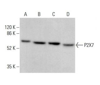
  • P2X7 Antibody (D-1) is recommended for detection of P2X7 of mouse, rat and human origin by WB, IP, IF and ELISA
  • Anti-P2X7 Antibody (D-1) is available conjugated to agarose for IP; HRP for WB, IHC(P) and ELISA; and to either phycoerythrin or FITC for IF, IHC(P) and FCM
  • also available conjugated to Alexa Fluor® 488, Alexa Fluor® 546, Alexa Fluor® 594 or Alexa Fluor® 647 for WB (RGB), IF, IHC(P) and FCM, and for use with RGB fluorescent imaging systems, such as iBright™ FL1000, FluorChem™, Typhoon, Azure and other comparable systems
  • also available conjugated to Alexa Fluor® 680 or Alexa Fluor® 790 for WB (NIR), IF and FCM; for use with Near-Infrared (NIR) detection systems, such as LI-COR®Odyssey®, iBright™ FL1000, FluorChem™, Typhoon, Azure and other comparable systems
  • m-IgG2a BP-HRP and m-IgGκ BP-HRP are the preferred secondary detection reagents for P2X7 Antibody (D-1) for WB applications. These reagents are now offered in bundles with P2X7 Antibody (D-1) (see ordering information below).
Gene Editing Promo Banner

QUICK LINKS

SEE ALSO...

P2X7 Antibody (D-1) is a mouse monoclonal IgG2a kappa light chain antibody that detects P2X7 of mouse, rat, and human origin by western blotting (WB), immunoprecipitation (IP), immunofluorescence (IF), and enzyme-linked immunosorbent assay (ELISA). P2X7 (D-1) antibody is available in both non-conjugated and various conjugated forms, including agarose, horseradish peroxidase (HRP), phycoerythrin (PE), fluorescein isothiocyanate (FITC), and multiple Alexa Fluor® conjugates. P2X7 is a member of the P2X receptor family, which consists of ligand-gated ion channels that facilitate calcium ion influx in response to extracellular ATP. P2X7 is predominantly located in the plasma membrane of various cell types, including neurons and immune cells, where P2X7 plays a crucial role in mediating synaptic transmission and inflammatory responses. P2X7 can trigger pro-inflammatory cytokine release and is implicated in processes such as necrosis and apoptosis when exposed to high ATP concentrations. Understanding P2X7 function and location is vital for elucidating P2X7′s role in various physiological and pathological conditions, including neurodegenerative diseases and chronic inflammation.

For Research Use Only. Not Intended for Diagnostic or Therapeutic Use.

Alexa Fluor® is a trademark of Molecular Probes Inc., OR., USA

LI-COR® and Odyssey® are registered trademarks of LI-COR Biosciences

P2X7 Antibody (D-1) References:

  1. Cytolytic P2X purinoceptors.  |  Di Virgilio, F., et al. 1998. Cell Death Differ. 5: 191-9. PMID: 10200464
  2. Allosteric modulation and accelerated resensitization of human P2X(3) receptors by cibacron blue.  |  Alexander, K., et al. 1999. J Pharmacol Exp Ther. 291: 1135-42. PMID: 10565834
  3. Inactivation of P2X2 purinoceptors by divalent cations.  |  Ding, S. and Sachs, F. 2000. J Physiol. 522 Pt 2: 199-214. PMID: 10639098
  4. Evidence for P2X(3), P2X(4), P2X(5) but not for P2X(7) containing purinergic receptors in Müller cells of the rat retina.  |  Jabs, R., et al. 2000. Brain Res Mol Brain Res. 76: 205-10. PMID: 10762695
  5. A natural dominant negative P2X1 receptor due to deletion of a single amino acid residue.  |  Oury, C., et al. 2000. J Biol Chem. 275: 22611-4. PMID: 10816552
  6. P2X receptors in sensory neurones.  |  Burnstock, G. 2000. Br J Anaesth. 84: 476-88. PMID: 10823099
  7. Pharmacology of cloned P2X receptors.  |  North, RA. and Surprenant, A. 2000. Annu Rev Pharmacol Toxicol. 40: 563-80. PMID: 10836147
  8. Function of the P2X7 receptor in hematopoiesis and leukemogenesis.  |  He, X., et al. 2021. Exp Hematol. 104: 40-47. PMID: 34687808
  9. Involvement of P2X7 receptors in chronic pain disorders.  |  Ren, WJ. and Illes, P. 2022. Purinergic Signal. 18: 83-92. PMID: 34799827
  10. The P2X7 receptor in mucosal adaptive immunity.  |  Grassi, F. and Marino, R. 2024. Purinergic Signal. 20: 9-19. PMID: 37067746
  11. Untangling Macropore Formation and Current Facilitation in P2X7.  |  Cevoli, F., et al. 2023. Int J Mol Sci. 24: PMID: 37446075
  12. The human P2x1 receptor: molecular cloning, tissue distribution, and localization to chromosome 17.  |  Longhurst, PA., et al. 1996. Biochim Biophys Acta. 1308: 185-8. PMID: 8809107

Ordering Information

Product NameCatalog #UNITPriceQtyFAVORITES

P2X7 Antibody (D-1)

sc-514962
200 µg/ml
$322.00

P2X7 Antibody (D-1): m-IgGκ BP-HRP Bundle

sc-524886
200 µg Ab, 40 µg BP
$361.00

P2X7 Antibody (D-1): m-IgG2a BP-HRP Bundle

sc-546176
200 µg Ab; 10 µg BP
$361.00

P2X7 Antibody (D-1) AC

sc-514962 AC
500 µg/ml, 25% agarose
$424.00

P2X7 Antibody (D-1) HRP

sc-514962 HRP
200 µg/ml
$322.00

P2X7 Antibody (D-1) FITC

sc-514962 FITC
200 µg/ml
$336.00

P2X7 Antibody (D-1) PE

sc-514962 PE
200 µg/ml
$349.00

P2X7 Antibody (D-1) Alexa Fluor® 488

sc-514962 AF488
200 µg/ml
$364.00

P2X7 Antibody (D-1) Alexa Fluor® 546

sc-514962 AF546
200 µg/ml
$364.00

P2X7 Antibody (D-1) Alexa Fluor® 594

sc-514962 AF594
200 µg/ml
$364.00

P2X7 Antibody (D-1) Alexa Fluor® 647

sc-514962 AF647
200 µg/ml
$364.00

P2X7 Antibody (D-1) Alexa Fluor® 680

sc-514962 AF680
200 µg/ml
$364.00

P2X7 Antibody (D-1) Alexa Fluor® 790

sc-514962 AF790
200 µg/ml
$364.00

P2X7 (D-1) Neutralizing Peptide

sc-514962 P
100 µg/0.5 ml
$69.00

使用这个抗体可以做大鼠脑片石蜡切片的免疫荧光吗?可以提供实验条件吗

Asked by: running
感谢您的提问。非常抱歉,P2X7 抗体 (D-1): sc-514962不推荐用于石蜡切片的免疫荧光实验,冰冻切片是推荐的。谢谢。 Thank you for your question. We are sorry to say that P2X7 Antibody (D-1): sc-514962 is not recommended for immunofluorescence experiments on paraffin sections, frozen sections are recommended . Thank you.
Answered by: Jiajun Z
Date published: 2024-06-19

为什们WB条带主要出现在55KDa

Asked by: lelay
感谢您的提问。您可以拨打电话:021-60936350,发送邮件到asia@scbio.cn,或者我们官网上的在线聊天,联系我们的亚洲技术支持团队,了解更多信息。 Thank you for your question. Please contact our Technical Service Department for more information. Our Asian Technical Service team is available at (+86) 021-60936350 or asia@scbio.cn or by live chat on our website.
Answered by: Technical Service SH
Date published: 2023-03-08

小鼠海马wb条带出现在55KDa 是否正常?

Asked by: lelay
感谢您的提问。您可以拨打电话:021-60936350,发送邮件到asia@scbio.cn,或者我们官网上的在线聊天,联系我们的亚洲技术支持团队,了解更多信息。 Thank you for your question. Please contact our Technical Service Department for more information. Our Asian Technical Service team is available at (+86) 021-60936350 or asia@scbio.cn or by live chat on our website.
Answered by: Technical Service SH
Date published: 2023-03-08

Buongiorno, è possibile avere un campione da provare? grazie

Asked by: elena91
Grazie per la tua domanda. Si prega di contattare il nostro reparto di assistenza tecnica (o il distributore locale) per ulteriori informazioni su come ricevere un FREE 10 µg sample.
Answered by: Tech Support Europe
Date published: 2021-10-14

Are you sure it recognizes an intracellular epitope as the description indicates? It appears to me that residues 81-106 of P2X7 are extracellular.

Asked by: Magdiel
Thank you for your inquiry. Anti-P2X7 Antibody (D-1): sc-514962 is raised against an epitope mapping between amino acids 81-106 of human origin. Amino acid 47-334 is located extracellular (https://www.uniprot.org/uniprot/Q99572#family_and_domains).sc-514962 is reocnize the extracellular rigon.
Answered by: Technical Support 9
Date published: 2021-07-14

I would like to know if the specificity of the antibody has been tested e.g. in a KO model.

Asked by: Nayana
Thank you for your question. The specificity of P2X7 Antibody (D-1): sc-514962 has been tested before the release of the antibody. If you are experiencing issues with this antibody, please contact our technical service for assistance.
Answered by: SCBT Heidelberg Support
Date published: 2019-01-06

我想用下你们的小样看看效果,货号sc-514962

Asked by: 张勇zhangyong
感谢您的提问。关于试用装的申请,欢迎通过:电话(+86) 021-60936350、邮箱asia@scbio.cn、或选择网页上Live Chat中的“中文支持”,联系我们的技术支持部门。 Thank you for your inquiry. For sample request, please contact our Asian Technical Service team at (+86) 021-60936350 or asia@scbio.cn or by live chat on our website.
Answered by: Technical Support 15
Date published: 2018-05-09

What application is the blocking peptide sc-514962 P appropriate for?

Asked by: Dr Ninau Qelp
Thank you for your question. The blocking peptide is intended for use as a negative control, by pre-adsorbing the mouse monoclonal antibody against the antigen. For full protocol details, please contact our Technical Services Department or view our online protocol here: https://www.scbt.com/scbt/resources/protocols/peptide-neutralization
Answered by: Technical Support
Date published: 2017-02-24
  • y_2026, m_4, d_29, h_11CST
  • bvseo_bulk, prod_bvqa, vn_bulk_3.0.43
  • cp_1, bvpage1
  • co_hasquestionsanswers, tq_8
  • loc_en_US, sid_514962, prod, sort_[SortEntry(order=LAST_APPROVED_ANSWER_SUBMISSION_TIME, direction=DESCENDING)]
  • clientName_scbt
  • bvseo_sdk, java_sdk, bvseo-4.0.0
  • CLOUD, getContent, 98ms
  • QUESTIONS, PRODUCT
Rated 5 out of 5 by from great figureThis product work well in WB at 1:800, liver cell,finally, I obtained clean and beautiful band !
Date published: 2017-11-27
Rated 5 out of 5 by from Results were great.85 kDa specific bands appear when they are diluted with WB 1/500 in HeLa cells. Many papers reported this antibody, and the quality is really good.
Date published: 2017-11-02
Rated 5 out of 5 by from The effect is very goodThis antibody is good for WB, I have a strong band and clean background. I think it's very useful.
Date published: 2017-10-27
Rated 5 out of 5 by from High sensitivityWe used P2X7 antibody got specific staining by cell lysates of human. Not only the stains very brightly but also the signal is powerful.
Date published: 2017-10-16
Rated 5 out of 5 by from It works great!I got a free sample and it worked great for western blot using rat brain proteins.
Date published: 2017-08-07
Rated 5 out of 5 by from effect is goodP2X7 antibody effect is good, immunohistochemical staining uniform, positive coloring significantly.
Date published: 2017-07-21
Rated 5 out of 5 by from Specific resultsWe have used it for WB analysis in spinal cord of rats: this antibody test sample was perfect, strong and very specific results!
Date published: 2017-06-29
Rated 5 out of 5 by from Popular antibodyVery good antibody WB detection P2X7 receptor analysis, welcomed by the laboratory people.
Date published: 2017-06-01
  • y_2026, m_4, d_29, h_11
  • bvseo_bulk, prod_bvrr, vn_bulk_3.0.43
  • cp_1, bvpage1
  • co_hasreviews, tv_0, tr_12
  • loc_en_US, sid_514962, prod, sort_[SortEntry(order=SUBMISSION_TIME, direction=DESCENDING)]
  • clientName_scbt
  • bvseo_sdk, java_sdk, bvseo-4.0.0
  • CLOUD, getReviews, 28ms
  • REVIEWS, PRODUCT
P2X7 Antibody (D-1) is rated 4.9 out of 5 by 12.
  • y_2026, m_4, d_29, h_11
  • bvseo_bulk, prod_bvrr, vn_bulk_3.0.43
  • cp_1, bvpage1
  • co_hasreviews, tv_0, tr_12
  • loc_en_US, sid_514962, prod, sort_[SortEntry(order=SUBMISSION_TIME, direction=DESCENDING)]
  • clientName_scbt
  • bvseo_sdk, java_sdk, bvseo-4.0.0
  • CLOUD, getAggregateRating, 115ms
  • REVIEWS, PRODUCT