")
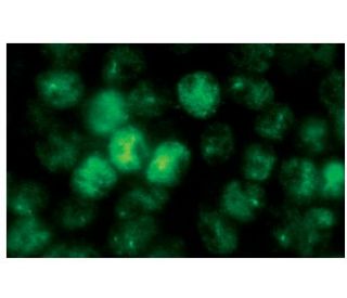
: sc-51690. Immunofluorescence staining of methanol-fixed EGF-treated A-431 cells showing nuclear localization.")
: sc-51690. Western blot analysis of p53 phosphorylation expression in untreated (A) and EGF treated (B) A-431 whole cell lysates.")
, untreated mouse p53 transfected: sc-125766 (B, E) and lambda protein phosphatase treated mouse p53 transfected: sc-125766 (C, F) 293T whole cell lysates. Antibodies tested include p-p53 Antibody (FP3.2): sc-51690 (A, B, C) and p53 (M-19): sc-1312 (D, E, F).")
p-p53 Antibody (FP3.2): sc-51690
- p-p53 Antibody (FP3.2) is a mouse monoclonal IgG1 κ, cited in 17 publications, provided at 100 µg/ml
- raised against a C-terminal phosphopeptide corresponding to amino acids Ser 392 of p53 of human origin
- recommended for detection of Ser 392 phosphorylated p53 of mouse, rat and human origin by WB, IP, IF, IHC(P) and ELISA; not recommended for detection of dephosphorylated p53
- m-IgG Fc BP-HRP and m-IgG1 BP-HRP are the preferred secondary detection reagents for p-p53 Antibody (FP3.2) for WB and IHC(P) applications. These reagents are now offered in bundles with p-p53 Antibody (FP3.2) (see ordering information below).

QUICK LINKS
SEE ALSO...
p-p53 Antibody (FP3.2) is a mouse monoclonal IgG1 kappa light chain antibody that detects p-p53, specifically Ser 392 phosphorylated p53, in mouse, rat, and human samples through various applications including western blotting (WB), immunoprecipitation (IP), immunofluorescence (IF), immunohistochemistry, and enzyme-linked immunosorbent assay (ELISA). p-p53 monoclonal antibody (FP3.2) is offered in a non-conjugated form, allowing for versatile experimental setups. p53 plays a critical role as a tumor suppressor, primarily functioning as a transcription factor that regulates the expression of genes involved in cell cycle control, apoptosis, and DNA repair in response to cellular stress. p53′s ability to localize to the nucleus is essential for function, as p53 binds to specific DNA sequences to activate target genes that promote growth arrest and programmed cell death. Notably, p53 undergoes various post-translational modifications, including phosphorylation, which can significantly influence activity and stability. Phosphorylation at Ser 392 enhances p53′s transcriptional activity and interaction with other regulatory proteins, thereby amplifying tumor-suppressive functions. Dysregulation of p53, often through mutations or aberrant modifications, is a common feature in many cancers, underscoring the importance of studying p53 for understanding tumor biology and developing targeted therapies. anti-p-p53 antibody (FP3.2) provides a valuable tool for studying the functional dynamics and regulatory mechanisms of p53 phosphorylation in both normal cellular processes and cancer research.
Ordering Information
| Product Name | Catalog # | UNIT | Price | Qty | FAVORITES | |
p-p53 Antibody (FP3.2) | sc-51690 | 100 µg/ml | $322.00 | |||
p-p53 Antibody (FP3.2): m-IgG Fc BP-HRP Bundle | sc-538943 | 100 µg Ab; 10 µg BP | $361.00 | |||
p-p53 Antibody (FP3.2): m-IgG1 BP-HRP Bundle | sc-541169 | 100 µg Ab; 20 µg BP | $361.00 |