
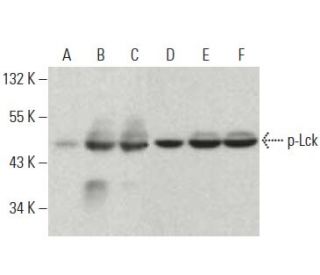
Anticorps p-Lck (pY505.4): sc-136184
- L'Anticorps p-Lck (pY505.4) est un monoclonal de souris IgG1 κ, cité dans 1 publications, fourni en 200 µg/ml
- élevé contre une courte séquence d'acides aminés contenant le Tyr 505 phosphorylé de Lck d'origine human.
- recommandé pour la détection de Tyr 505 phosphorylated Lck d'origine mouse, rat et human par WB et IP
- Le m-IgGκ BP-HRP est le réactif de détection secondaire préféré pour l'anticorps p-Lck (pY505.4) pour les applications WB. Ce réactif est désormais proposé en association avec l'anticorps p-Lck (pY505.4)(voir les informations relatives à la commande ci-dessous). Pour d'autres conjugués m-IgGκ BP, voir notre liste complète de protéines de liaison à l'IgG de souris.

ACCÈS RAPIDE AUX LIENS
VOIR ÉGALEMENT...
La famille des gènes Src de protéines tyrosine kinases associées à la membrane comprend c-Src, c-Yes, Fyn, Lck, Hck, Lyn, Blk et c-Fgr. Le gène humain Lck code pour une tyrosine kinase spécifique des lymphocytes, désignée pp56 Lck. Le réarrangement et la surexpression du gène Lck se produisent dans certains lymphomes murins. Les lymphomes et les neuroblastomes humains présentent fréquemment des anomalies chromosomiques dans un site du génome qui contient le gène Lck. Les cellules T au repos contiennent des niveaux élevés de protéine et d'ARNm Lck, qui diminuent tous deux lors de l'activation des cellules T. L'expression de Lck peut contribuer à la pathogénicité de la maladie. L'expression de Lck peut contribuer à la pathogenèse de certains types de maladies néoplasiques. Csk phosphoryle Lck sur Tyr 505. Cette phosphorylation supprime l'activité catalytique de Lck dans certaines conditions. La phosphorylation de Lck sur Tyr 394 stimule l'activité de Lck indépendamment de l'état de phosphorylation de Tyr 505. En réponse à la liaison du récepteur de l'antigène des cellules T et à la signalisation qui s'ensuit, Lck subit une phosphorylation sur des résidus qui comprennent Tyr 192, Ser 59 et Ser 194.
Alexa Fluor® est une marque déposée de Molecular Probes Inc., OR., USA
LI-COR® et Odyssey® sont marques déposées de LI-COR Biosciences
Anticorps p-Lck (pY505.4) Références:
- Caractérisation des sites in vivo de phosphorylation de la sérine sur Lck, identifiant la sérine 59 comme un site de phosphorylation mitotique. | Kesavan, KP., et al. 2002. J Biol Chem. 277: 14666-73. PMID: 11847223
- La tyrosine kinase humaine p50csk phosphoryle p56lck à Tyr-505 et régule à la baisse son activité catalytique. | Bergman, M., et al. 1992. EMBO J. 11: 2919-24. PMID: 1639064
- Expression et interactions de la famille Src des tyrosines protéines kinases dans les lymphocytes T. | Bolen, JB., et al. 1991. Adv Cancer Res. 57: 103-49. PMID: 1950702
- Un gène de protéine-tyrosine kinase spécifique des lymphocytes est réarrangé et surexprimé dans le lymphome murin à cellules T LSTRA. | Marth, JD., et al. 1985. Cell. 43: 393-404. PMID: 2416464
- Régulation de pp56lck pendant l'activation des cellules T: implications fonctionnelles pour les protéines tyrosine kinases de type src. | Marth, JD., et al. 1987. EMBO J. 6: 2727-34. PMID: 3119327
- Activation de p56lck par p72syk par association physique et phosphorylation de la tyrosine N-terminale. | Couture, C., et al. 1994. Mol Cell Biol. 14: 5249-58. PMID: 7518561
- La stimulation de la phosphorylation de Tyr394 par le peroxyde d'hydrogène réactive les formes biologiquement inactives de Lck, non liées à la membrane. | Yurchak, LK., et al. 1996. J Biol Chem. 271: 12549-54. PMID: 8647864
- Régulation du domaine SH2 de Lck par la phosphorylation de la tyrosine. | Couture, C., et al. 1996. J Biol Chem. 271: 24880-4. PMID: 8798764
- Base structurelle de l'activation de la kinase lymphocytaire humaine Lck par phosphorylation de la tyrosine. | Yamaguchi, H. and Hendrickson, WA. 1996. Nature. 384: 484-9. PMID: 8945479
- Dans les cellules exposées au peroxyde d'hydrogène, la forme activée de la tyrosine protéine kinase Lck est phosphorylée à la fois sur Tyr-394 et Tyr-505. | Hardwick, JS. and Sefton, BM. 1997. J Biol Chem. 272: 25429-32. PMID: 9325251
Informations pour la commande
| Nom du produit | Ref. Catalogue | COND. | Prix HT | QTÉ | Favoris | |
Anticorps p-Lck (pY505.4) | sc-136184 | 200 µg/ml | $322.00 | |||
p-Lck (pY505.4): m-IgGκ BP-HRP Kit | sc-521339 | 200 µg Ab, 40 µg BP | $361.00 |