")
OAT Antibody (A-12): sc-374243
- OAT Antibody (A-12) is a mouse monoclonal IgG2a κ, cited in 5 publications, provided at 200 µg/ml
- raised against amino acids 96-230 mapping within an internal region of OAT of human origin
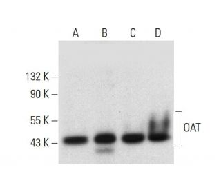
- Anti-OAT Antibody (A-12) is recommended for detection of hepatic and renal forms of ornithine aminotransferase of mouse, rat and human origin by WB, IP, IF, IHC(P) and ELISA
- Anti-OAT Antibody (A-12) is available conjugated to agarose for IP; HRP for WB, IHC(P) and ELISA; and to either phycoerythrin or FITC for IF, IHC(P) and FCM
- also available conjugated to Alexa Fluor® 488, Alexa Fluor® 546, Alexa Fluor® 594 or Alexa Fluor® 647 for WB (RGB), IF, IHC(P) and FCM, and for use with RGB fluorescent imaging systems, such as iBright™ FL1000, FluorChem™, Typhoon, Azure and other comparable systems
- also available conjugated to Alexa Fluor® 680 or Alexa Fluor® 790 for WB (NIR), IF and FCM; for use with Near-Infrared (NIR) detection systems, such as LI-COR®Odyssey®, iBright™ FL1000, FluorChem™, Typhoon, Azure and other comparable systems
- m-IgG Fc BP-HRP is the preferred secondary detection reagent for OAT Antibody (A-12) for WB and IHC(P) applications. This reagent is now offered in a bundle with OAT Antibody (A-12) (see ordering information below).

QUICK LINKS
OAT Antibody (A-12) is a mouse monoclonal IgG2a kappa light chain antibody that detects ornithine aminotransferase (OAT) in mouse, rat, and human samples through applications such as western blotting (WB), immunoprecipitation (IP), immunofluorescence (IF), immunohistochemistry with paraffin-embedded sections (IHCP), and enzyme-linked immunosorbent assay (ELISA). OAT antibody (A-12) is available in both non-conjugated and various conjugated forms, including agarose, horseradish peroxidase (HRP), phycoerythrin (PE), fluorescein isothiocyanate (FITC), and multiple Alexa Fluor® conjugates. Ornithine aminotransferase (OAT) is a crucial enzyme composed of 439 amino acids, encoded by the human gene OAT, and belongs to the class III pyridoxal-phosphate-dependent aminotransferase family. OAT is primarily located in the mitochondrial matrix, where OAT functions as a homotetramer, catalyzing the conversion of ornithine to other metabolites, which is vital for the urea cycle and amino acid metabolism. Proper functioning of OAT is essential, as deficiencies can lead to ornithinemia, a condition associated with gyrate atrophy of the choroid and retina, characterized by progressive vision loss and retinal degeneration. The hepatic form of OAT is generated by the cleavage of a 25 amino acid transit peptide from the N-terminus of its precursor, while the renal form arises from the cleavage of a 35 amino acid transit peptide, highlighting the importance of post-translational modifications in determining OAT′s functional roles in different tissues.
Alexa Fluor® is a trademark of Molecular Probes Inc., OR., USA
LI-COR® and Odyssey® are registered trademarks of LI-COR Biosciences
OAT Antibody (A-12) References:
- Somatic versus germline mutation processes at minisatellite CEB1 (D2S90) in humans and transgenic mice. | Buard, J., et al. 2000. Genomics. 65: 95-103. PMID: 10783256
- Strand-separating conformational polymorphism analysis: efficacy of detection of point mutations in the human ornithine delta-aminotransferase gene. | Michaud, J., et al. 1992. Genomics. 13: 389-94. PMID: 1612597
- Ornithine aminotransferase deficiency: diagnostic difficulties in neonatal presentation. | Cleary, MA., et al. 2005. J Inherit Metab Dis. 28: 673-9. PMID: 16151897
- Dietary compliance in ornithine aminotransferase deficiency. | Santos, L., et al. 2006. J Inherit Metab Dis. 29: 240. PMID: 16601905
- Cellular response to 5-fluorouracil (5-FU) in 5-FU-resistant colon cancer cell lines during treatment and recovery. | De Angelis, PM., et al. 2006. Mol Cancer. 5: 20. PMID: 16709241
- Ornithine deficiency in the arginase double knockout mouse. | Deignan, JL., et al. 2006. Mol Genet Metab. 89: 87-96. PMID: 16753325
- Molecular pathology of gyrate atrophy of the choroid and retina due to ornithine aminotransferase deficiency. | Ramesh, V., et al. 1991. Mol Biol Med. 8: 81-93. PMID: 1682785
- Human ornithine aminotransferase complexed with L-canaline and gabaculine: structural basis for substrate recognition. | Shah, SA., et al. 1997. Structure. 5: 1067-75. PMID: 9309222
Ordering Information
| Product Name | Catalog # | UNIT | Price | Qty | FAVORITES | |
OAT Antibody (A-12) | sc-374243 | 200 µg/ml | $322.00 | |||
OAT Antibody (A-12): m-IgG Fc BP-HRP Bundle | sc-525944 | 200 µg Ab; 10 µg BP | $361.00 | |||
OAT Antibody (A-12) AC | sc-374243 AC | 500 µg/ml, 25% agarose | $424.00 | |||
OAT Antibody (A-12) HRP | sc-374243 HRP | 200 µg/ml | $322.00 | |||
OAT Antibody (A-12) FITC | sc-374243 FITC | 200 µg/ml | $336.00 | |||
OAT Antibody (A-12) PE | sc-374243 PE | 200 µg/ml | $349.00 | |||
OAT Antibody (A-12) Alexa Fluor® 488 | sc-374243 AF488 | 200 µg/ml | $364.00 | |||
OAT Antibody (A-12) Alexa Fluor® 546 | sc-374243 AF546 | 200 µg/ml | $364.00 | |||
OAT Antibody (A-12) Alexa Fluor® 594 | sc-374243 AF594 | 200 µg/ml | $364.00 | |||
OAT Antibody (A-12) Alexa Fluor® 647 | sc-374243 AF647 | 200 µg/ml | $364.00 | |||
OAT Antibody (A-12) Alexa Fluor® 680 | sc-374243 AF680 | 200 µg/ml | $364.00 | |||
OAT Antibody (A-12) Alexa Fluor® 790 | sc-374243 AF790 | 200 µg/ml | $364.00 |