Date published: 2026-4-4

1-800-457-3801

SCBT Portrait Logo
Seach Input

MAP-2 Antibody (A-8): sc-74422

5.0(6)
Write a reviewAsk a question

Datasheets
  • MAP-2 Antibody (A-8) is a mouse monoclonal IgG2a κ, cited in 13 publications, provided at 200 µg/ml
  • raised against amino acids 1-300 of MAP-2 of human origin
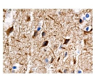
  • recommended for detection of MAP-2 of mouse, rat and human origin by WB, IP, IF, IHC(P) and ELISA
  • See MAP-2 (A-4): sc-74421 for MAP-2 antibody conjugates, including AC, HRP, FITC, PE, Alexa Fluor® 488, 594, 647, 680 and 790.
  • m-IgG2a BP-HRP is the preferred secondary detection reagent for MAP-2 Antibody (A-8) for WB and IHC(P) applications. This reagent is now offered in a bundle with MAP-2 Antibody (A-8) (see ordering information below).
Gene Editing Promo Banner

QUICK LINKS

SEE ALSO...

MAP-2 Antibody (A-8) is a mouse monoclonal IgG2a antibody that detects MAP-2 in mouse, rat, and human samples through various applications including western blotting (WB), immunoprecipitation (IP), immunofluorescence (IF), immunohistochemistry with paraffin-embedded sections (IHCP), and enzyme-linked immunosorbent assay (ELISA). MAP-2 is a crucial microtubule-associated protein that plays a significant role in stabilizing microtubules and facilitating their assembly, which is essential for maintaining the structural integrity of the cytoskeleton in neurons. MAP-2 is predominantly located in the dendrites of neurons, where MAP-2 contributes to the formation of dendritic spines and the overall architecture of the neuronal network. Proper localization of MAP-2 is vital for neuronal function, as MAP-2 influences synaptic plasticity and signal transduction pathways that are critical for learning and memory. Additionally, MAP-2 is subject to various post-translational modifications, including phosphorylation, which can modulate MAP-2′s activity and interactions with other proteins, further underscoring MAP-2′s importance in cellular dynamics and neurodevelopment. Anti-MAP-2 antibody (A-8) is an invaluable tool for researchers studying cytoskeletal dynamics and neuronal architecture, providing insights into the molecular mechanisms underlying neurodegenerative diseases and other neurological disorders.

For Research Use Only. Not Intended for Diagnostic or Therapeutic Use.

Ordering Information

Product NameCatalog #UNITPriceQtyFAVORITES

MAP-2 Antibody (A-8)

sc-74422
200 µg/ml
$322.00

MAP-2 Antibody (A-8): m-IgG2a BP-HRP Bundle

sc-546009
200 µg Ab; 10 µg BP
$361.00

I am using MAP-2 (A-8): sc-74422 monoclonal antibody to stain paraffin embedded tissue. Do you recommend antigen retrieval when using this antibody?

Asked by: AbPolly
We recommend performing antigen retrieval with sodium citrate buffer (pH 6) and heat. The full protocol can be found here: https://www.scbt.com/scbt/resources/protocols/immunoperoxidase-staining
Answered by: Technical Support
Date published: 2020-08-11
  • y_2026, m_3, d_26, h_7CST
  • bvseo_bulk, prod_bvqa, vn_bulk_3.0.43
  • cp_1, bvpage1
  • co_hasquestionsanswers, tq_1
  • loc_en_US, sid_74422, prod, sort_[SortEntry(order=LAST_APPROVED_ANSWER_SUBMISSION_TIME, direction=DESCENDING)]
  • clientName_scbt
  • bvseo_sdk, java_sdk, bvseo-4.0.0
  • CLOUD, getContent, 97ms
  • QUESTIONS, PRODUCT
Rated 5 out of 5 by from observed target bandRat brian tissue extracts (50ug),1:800 diluted the primary, and 1:5000 diluted the secondary,The WB result is expected bands at 280kDa.
Date published: 2017-07-02
Rated 5 out of 5 by from worked good qualityI tried different antibody dilutions and to optimize it, but I always get the same (very strong) bands from 1:500 to 1:800 (Hela cell).
Date published: 2017-07-01
Rated 5 out of 5 by from Positive signal in WB in mouse transfectedPositive signal in WB in mouse transfected 293T whole cell lysates. -SCBT QC
Date published: 2015-03-19
Rated 5 out of 5 by from Excellent immunoperoxidase neuropil and cytoplasmicExcellent immunoperoxidase neuropil and cytoplasmic staining in formalin fixed, paraffin-embedded human brain tissue. -SCBT QC
Date published: 2015-03-16
Rated 5 out of 5 by from Robust signal in WB in human transfectedRobust signal in WB in human transfected 293T whole cell lysates. -SCBT QC
Date published: 2015-01-10
Rated 5 out of 5 by from Worked well in WB in mouse and rat brainWorked well in WB in mouse and rat brain tissue extracts. -SCBT QC
Date published: 2015-01-08
  • y_2026, m_3, d_26, h_7
  • bvseo_bulk, prod_bvrr, vn_bulk_3.0.43
  • cp_1, bvpage1
  • co_hasreviews, tv_0, tr_6
  • loc_en_US, sid_74422, prod, sort_[SortEntry(order=SUBMISSION_TIME, direction=DESCENDING)]
  • clientName_scbt
  • bvseo_sdk, java_sdk, bvseo-4.0.0
  • CLOUD, getReviews, 7ms
  • REVIEWS, PRODUCT
MAP-2 Antibody (A-8) is rated 5.0 out of 5 by 6.
  • y_2026, m_3, d_26, h_7
  • bvseo_bulk, prod_bvrr, vn_bulk_3.0.43
  • cp_1, bvpage1
  • co_hasreviews, tv_0, tr_6
  • loc_en_US, sid_74422, prod, sort_[SortEntry(order=SUBMISSION_TIME, direction=DESCENDING)]
  • clientName_scbt
  • bvseo_sdk, java_sdk, bvseo-4.0.0
  • CLOUD, getAggregateRating, 94ms
  • REVIEWS, PRODUCT